


Cross section of demineralized premolar (100X)
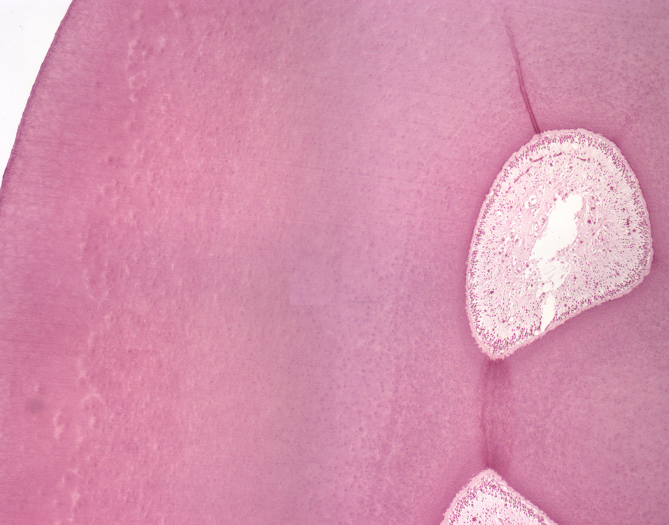
Areas of interglobular dentine are seen some distance from the junction between the dentine and the enamel. These areas of dentine represent unmineralized dentine. The interglobular dentine stains lighter than the dentine, more like the predentine. The dentinal tubules are visible throughout the dentine.
The odontoblasts in the pulp are easily spotted as a "ring of cells" covering the total circumference of the pulp cavity only to be separated from the dentine by the lighter colored predentine. In addition, you can see the same structures in the pulp as in the longitudinal section. Try to compare the two images and bear in mind that this is a cross section and the other is a longitudinal section.