


Longitudinal section through a demineralized premolar (40X)
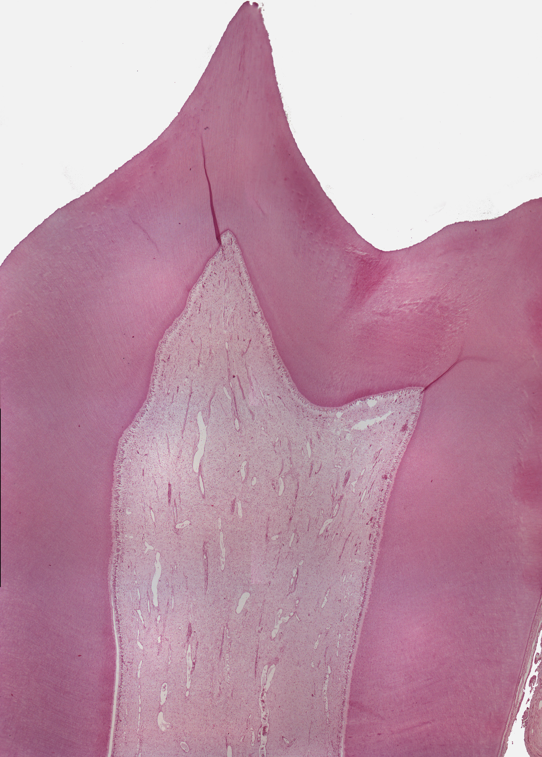
This is a histology image of a longitudinal section through a premolar. The enamel is missing due to tissue preparatory processes. The dentine in the root is covered by a thin layer of cementum. Magnify the preselected areas for further detail. The pulp can be seen in the middle below the dentine.