


Dentine and pulp in a demineralized cross section of a premolar (400X)
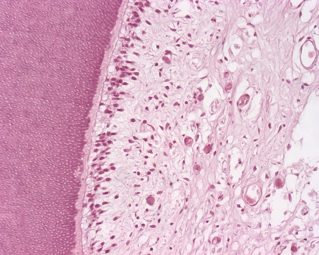
This image shows a few details from one of the pulp horns. The dentine is "jam packed" with cross sectioned dentintubuli. Predentine is visible adjacent to the odontoblast layer. Even further in you can see the cell free zone, the cell rich zone and finally the main body of pulp tissue that for the most part contains connective tissue.
If you've studied the longitudinal section of a premolar, you find that most blood vessels and nerves of the pulp are cut longitudinally, while in this section, you will see that most blood vessels and nerves are cut across.