


Cross section of a premolar showing the pulp and dentin (40X)
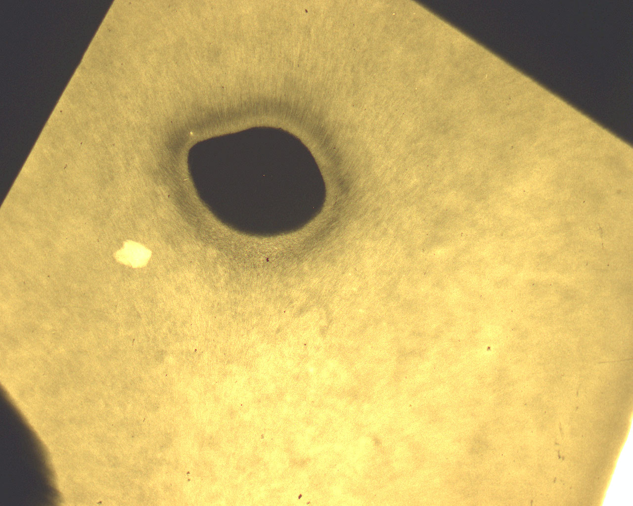
This is a microradiogram showing a cross section of a premolar. The image focuses on the circumpulpal dentine. There is also a longitudinal section of two premolars.
Do remember that this is a negative image meaning that light areas in an x-ray are highly mineralized and vice versa.
The pulp is centered in the image (lat: pulpa). The dentine circumvents the pulp. The dentine displays primarily a light sone closest to the pulp, then a darker zone, then the main body of the dentine.
Interglobular dentine can be seen in another cross section of a premolar.