


Microradiogram of a cross section of a premolar (40X)
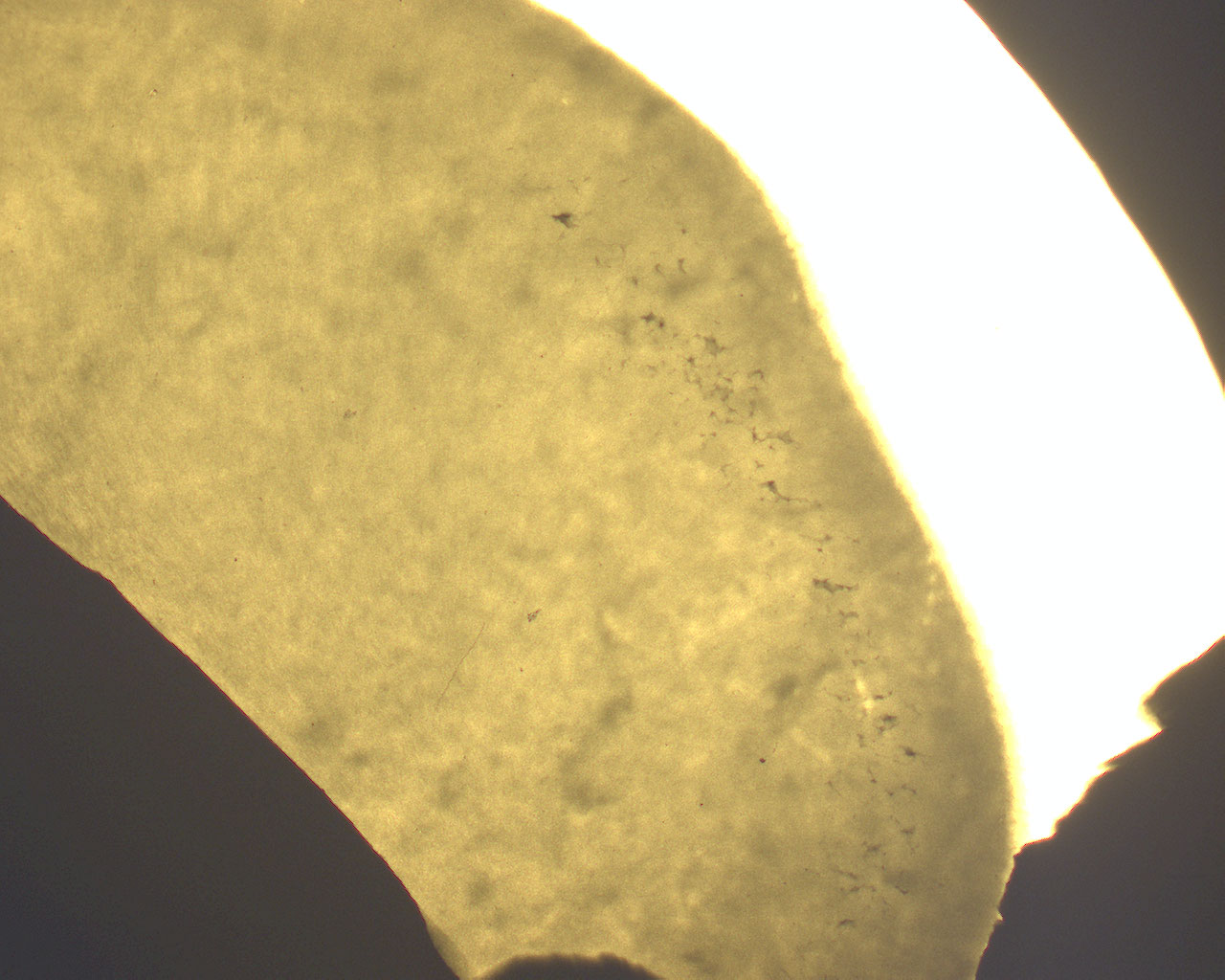
This is a microradiogram showing a cross section of a premolar and we're going to take a look at the dentine close to the enamel in this section. There's also a microradiogram of a longitudinal section of a premolar.
Remember that this is a negative image and the lighter areas represent tissue with high grade of mineralization and vice versa.
This image shows the dentine and the enamel. Close to the enamel, there's a region with interglobular dentine that is darker than the surrounding dentine and take on a star like appearance.