


Anterior eye with cornea, sclera and iris (40X)
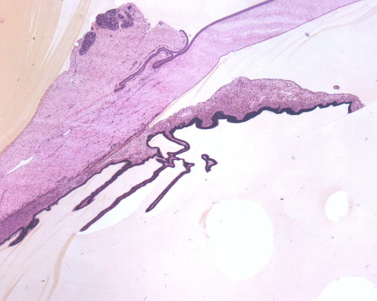
This is a section of the eye. In this series of pictures from this part of the eye, we concentrate on it's anterior structures. For practical reasons, one can divide the eye into
- an outer fibrous layer consisting of the cornea and the sclera
- a middle layer made up of the choroid, iris and vasculature
- an inner layer constituted by the retina (not visible in this image)
As in the other section of the anterior eye, you can see the outer and middle layers.
The hollow areas of the eye that separates the anterior parts of the eye is called camera anterior and camera posterior.
The sclera is a continuation of the cornea and consists of fibroblasts, a few elastic fibers and bundles of collagen of which all run parallel to the surface of the eye.