


Retina of the eye (40X)
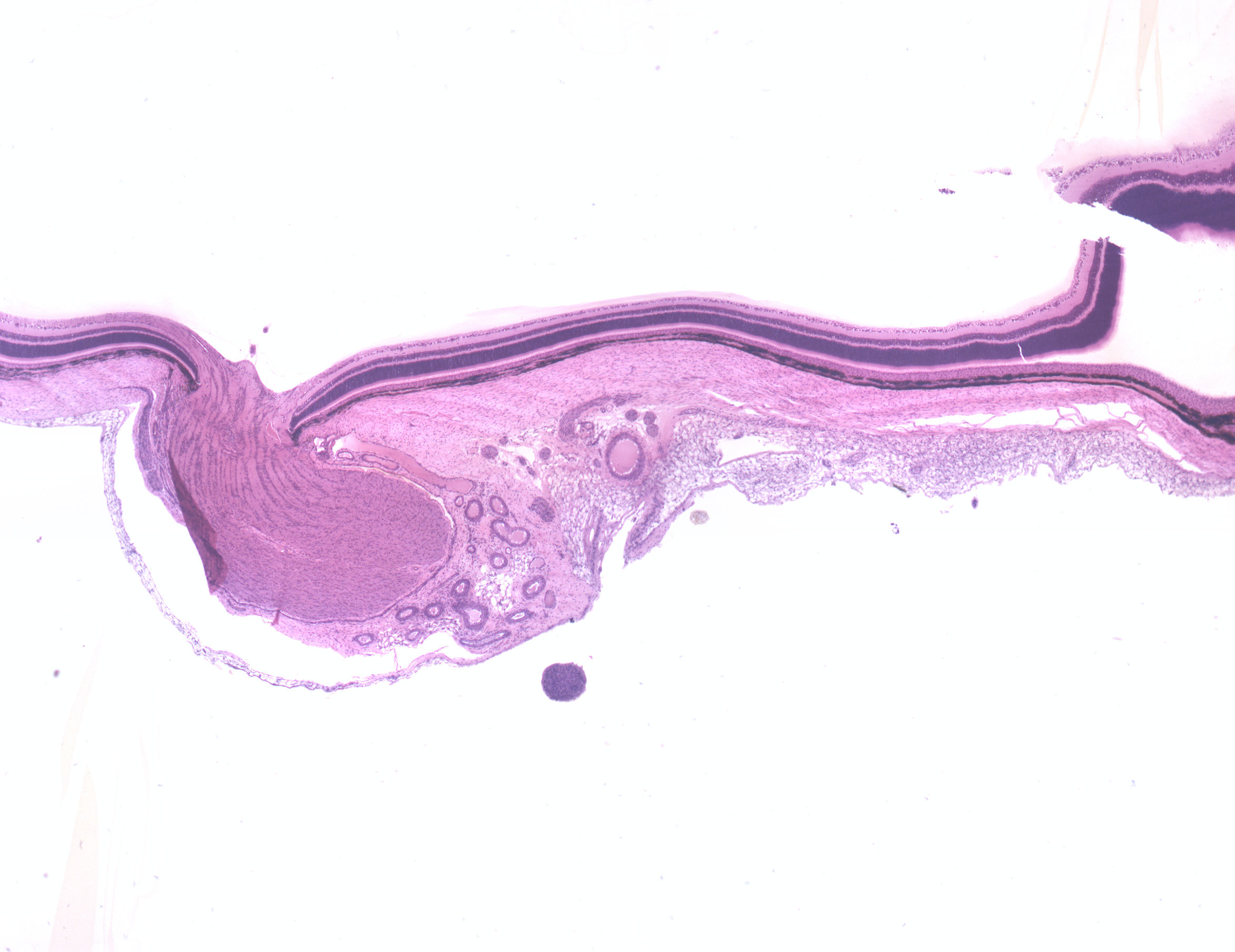
This is a section from the posterior part of the eye. For practical reasons, one can divide the eye into three layers;
- an outer fibrous layer consisting of the cornea and sclera
- a middle layer made up of the choroid, iris and vasculature
- an inner layer constituted by the retina (seen here).
Tissues from all three layers can be seen in the picture.
The sclera is a continuation of the cornea and consists of fibroblasts, a few elastic fibers and bundles of collagen of which all run parallel to the surface of the eye. Choroid is the dark membrane seen beween the retina and the sclera. The retina covers the inside of the eye. At this magnification, you can already see the retinas layered structure.
There are two land marks on the posterior wall of the retina: makula lutea and the optic papilla. Makula lutea (not seen in this image) is a small depression in the retina and contains only photo receptors of the coned type. This is where we humans have our "sharp vision". The optic papilla (lat: punctum caecum) is seen in the picture. This is where the optic nerve leaves the eye for the brain. There are no photo receptors at this area, that's why it's called the blind spot.