


Cornea and iris (100X)
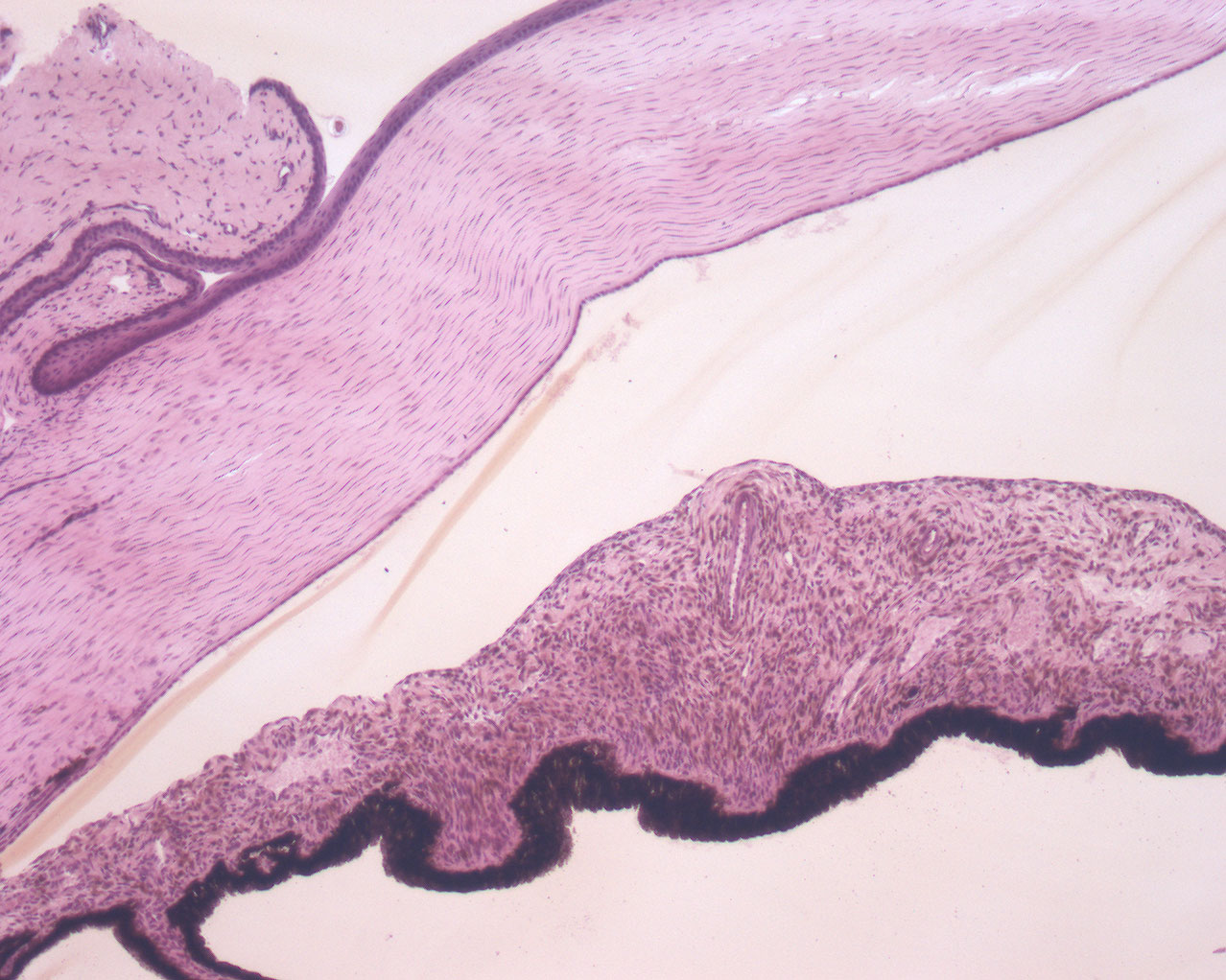
The cornea is covered by stratified squamous epithelium at the surface facing the eye lid (anterior part, also called the epithelium anterius cornae). On the opposite side the epithelial cells are cubic in shape. The cornea consists of parallel bundles of collagen. You can see keratinocytes interspersed in between these bundles.
The iris is the part of the eye that is colored macroscopically (blue, brown or green) and contains circular fibers that constitute the pupil. The iris is covered by the choroid posteriorly.