


Anterior eye with cornea and eyelid (40X)
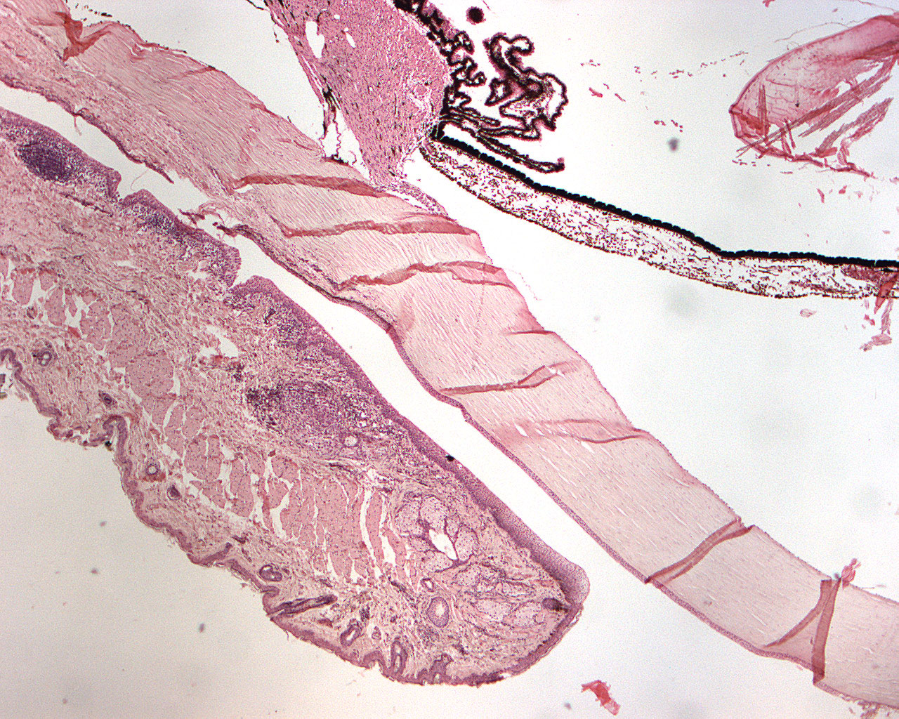
This section is from the anterior part of eye. You can see the
- cornea
- lens
- iris
- eye lid
- sclera
The tissue itself is not very well preserved and the lens is quite damaged. But a hair follicles, sweat- and tarsal glands are seen in the palpebra (eye lid).
The sclera is the outer fibrous layer of the eye. It is the "white stuff" around the pupil in the eye. The choroid is the dark membrane you can see below the sclera. It is highly vascularized. The iris is the colored part of the eye (blue, brown, green). It encircles the pupil.
Check the other section of the anterior eye for a more comprehensive explanation of anatomical structures.