


Cross section of a bone (40X)
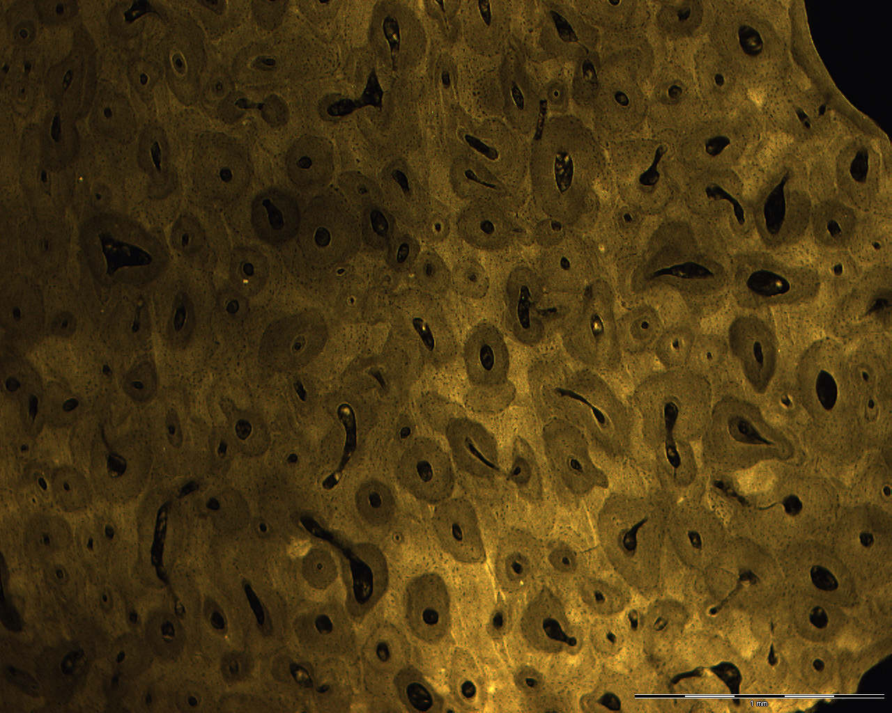
This is a microradiogram of a cross section of a compact bone. There is a longitudinal section of bone as well.
A microradiogram is a small x-ray of a tissue sample. You can then use the x-ray in a microscope for further study. That's what you see in the image.
The lighter areas are the ones which hold the highest portion of minerals. These areas are lighter because less radiation is let through to stain the x-ray film (the minerals block the x-rays).
The darker areas in the section are either Haversian- or Volkmann's canals.