


Longitudinal section through a hollow bone (40X)
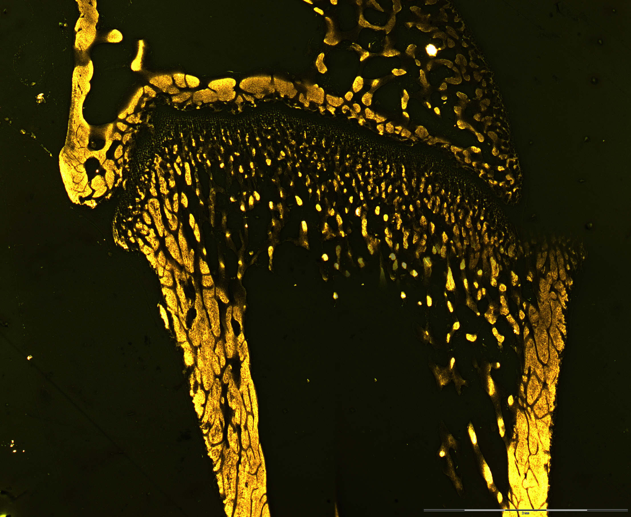
This is a microradiogram of a longitudinal section of a bone . There is a cross section of bone as well. The areas that appear lighter are the ones that are mineralized and block the x-rays an leave a "light shadow" on the x-ray film. The compact bone (cortical bone) is seen as lighter areas at both sides of the "black" bone marrow.
A large dark area is seen at the center of the image only to be phalanxed by two lighter yellow areas that represent the compact bone (cortical bone). The dark area is the bone marrow and the cavity that it occupies in a bone can be called a marrow cavity.
This longitudinal section is showing both the epi- and diaphysis, and the primary issificationcenter. It is impossible to view detail of it in a microradiogram. See if you can find any similarities in the H+E stained longitudinal section of a hollow bone.