
Ductus deferens (400X)
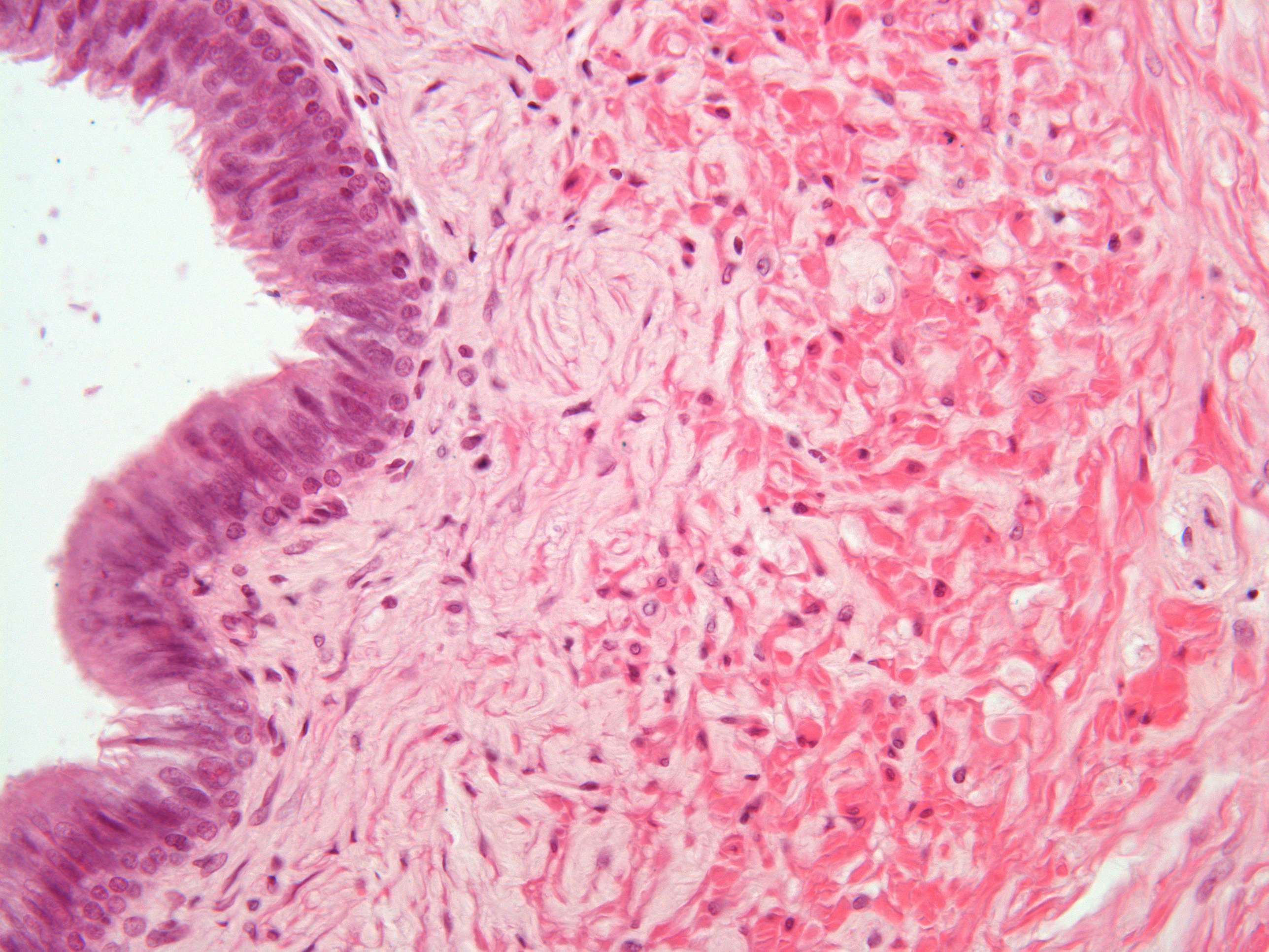
In this high power picture you can study details of the innermost layers of the ductus deferens (vas deferens). The epithelium is pseudostratified columnar. Some of the cells are equipped with sterocilia (like the epithelial cells in the ductus epididymidis). You can also easily distinguish the lamina propria and the innermost, longitudinal layer of smooth muscle cells (most cells are cut transversely).