
Ductus deferens (40X)
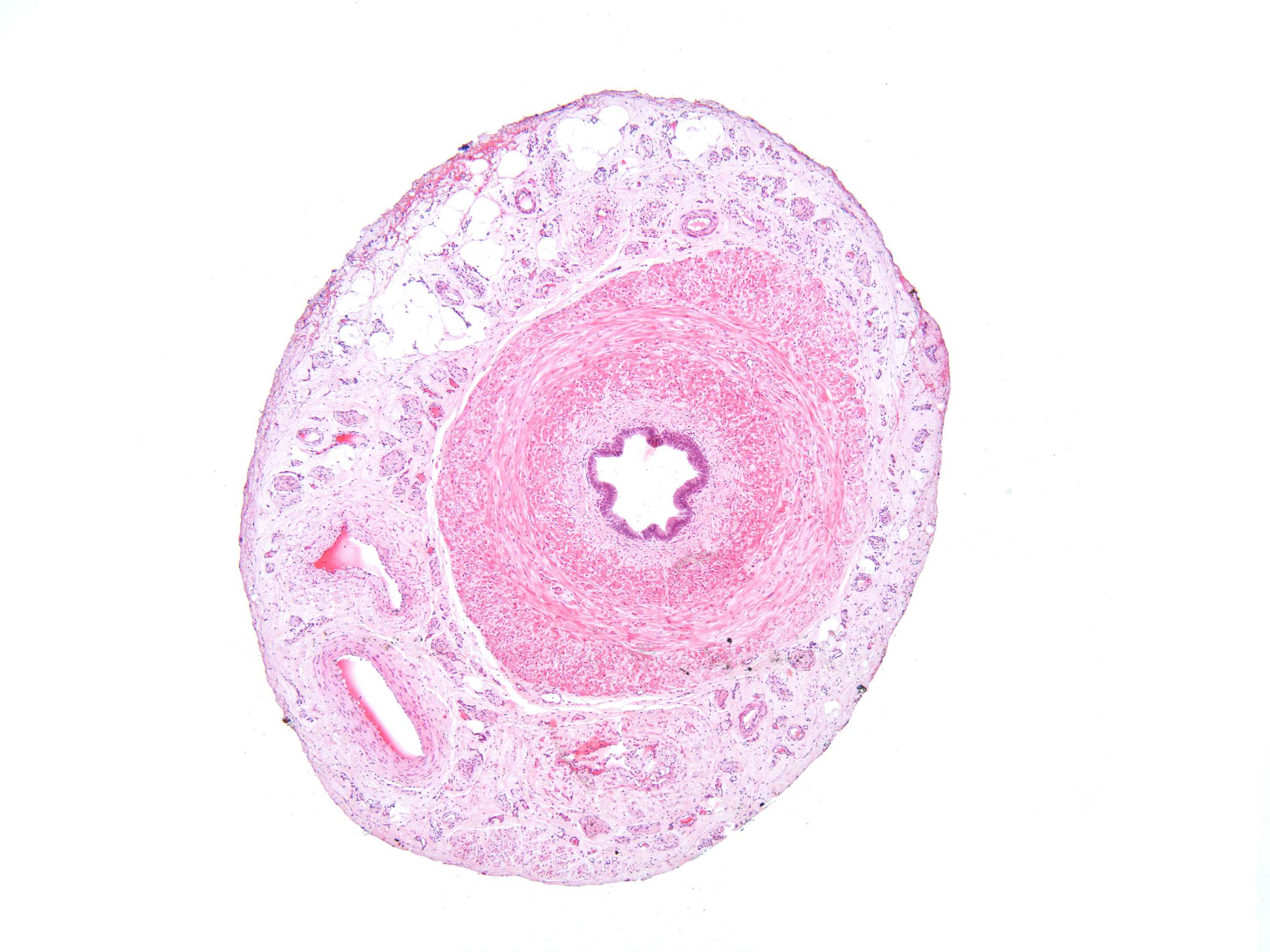
This is a cross section of the ductus deferens (vas deferens). The lumen is small and covered by purple epithelium facing the lumen. Surrounding the epithelium you see a lightly colored pink layer. This is the lamina propria. The thick, darker colored layer outside the lamina propria is made up of smooth muscle. We've labeled it tunica muscularis. The outermost layer of connective tissue is the tunica adventitia.