
Ductus deferens (100X)
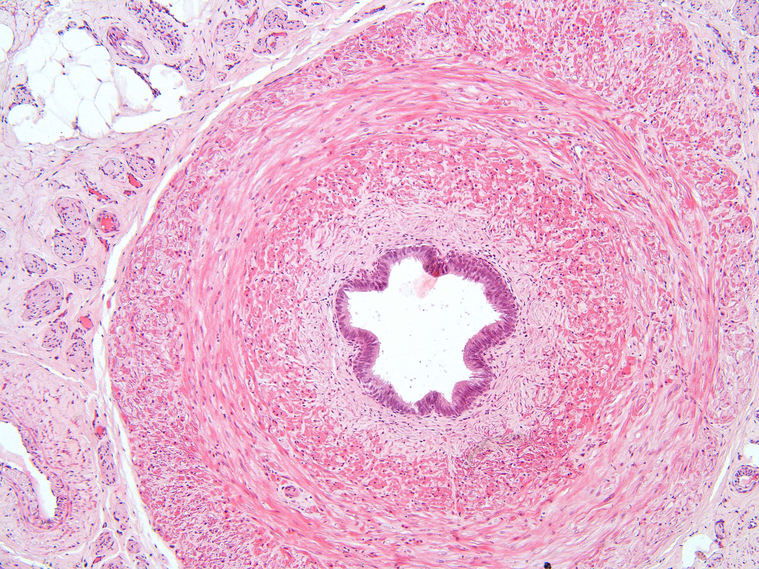
This magnified image shows the same structures as in the previous picture, but at this magnification you can see that the muscle layer can be divided into three different layers, an inner and outer longitudinal layer, separated by a circular layer.
Many nerve fascicles and veins lie close to the outer muscular layer. The nerve fascicles carry sensory fibers and many sympathetic postganglionic fibers (controlling peristaltic movements of the smooth muscles during ejaculation).