
Testis (40X)
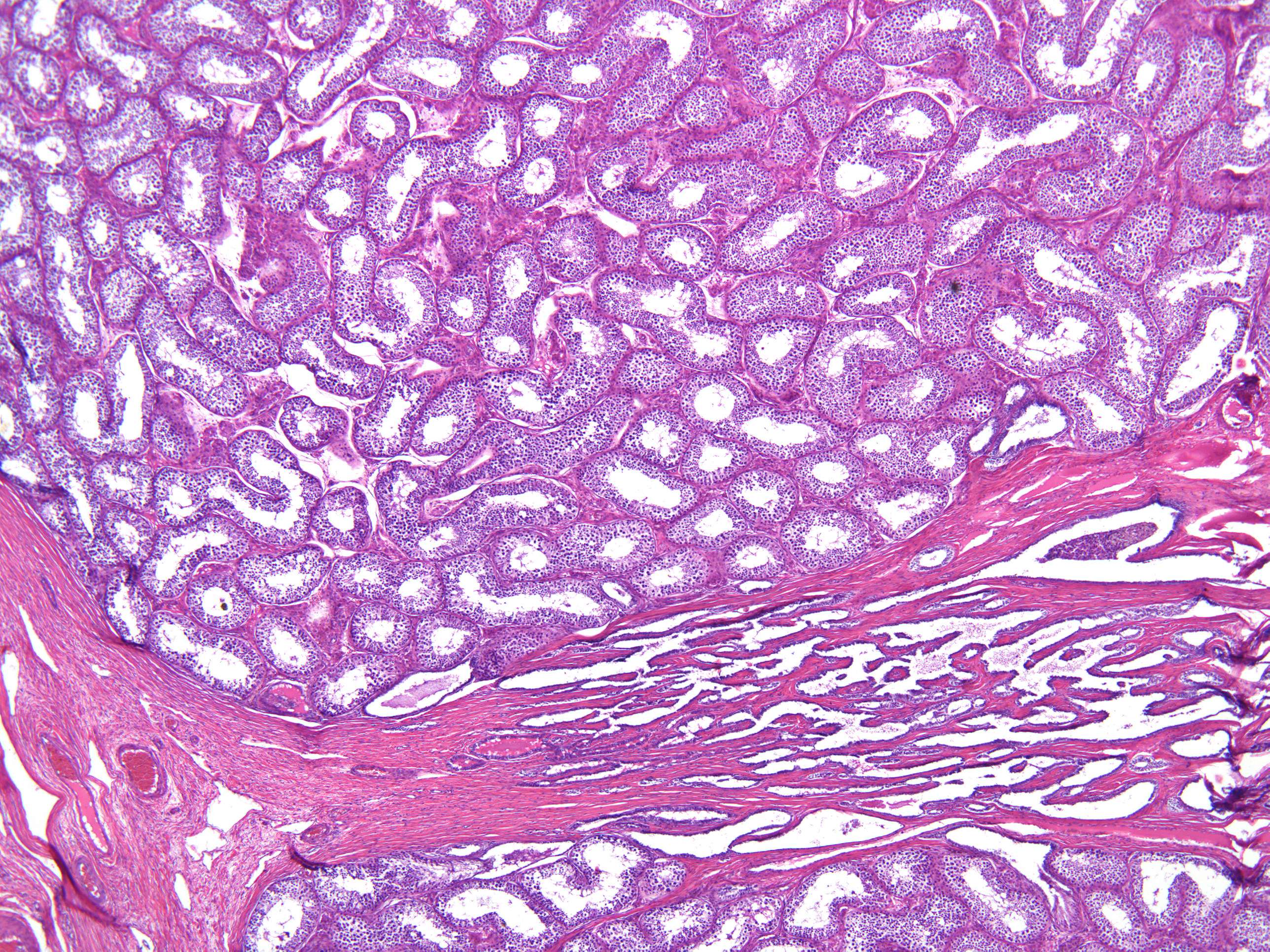
In this medium-power photomicrograph you see many bluish circular structures with a lumen in the middle. They are called tubuli seminiferi contorti (singular: tubulus seminiferous contortus) and this is where the magic happens: the production of the sperms (spermatozoa).
The fibrous structure seen at the bottom 1/3 of this image is the rete testis and is a meshwork of tubules that transports sperm from the tubuli seminiferi to the epididymis.
The space between the tubules is the interstitium and in this space is occupied by hormone producing cells the are more easily seen at higher magnification.