
Testis (12.5X)
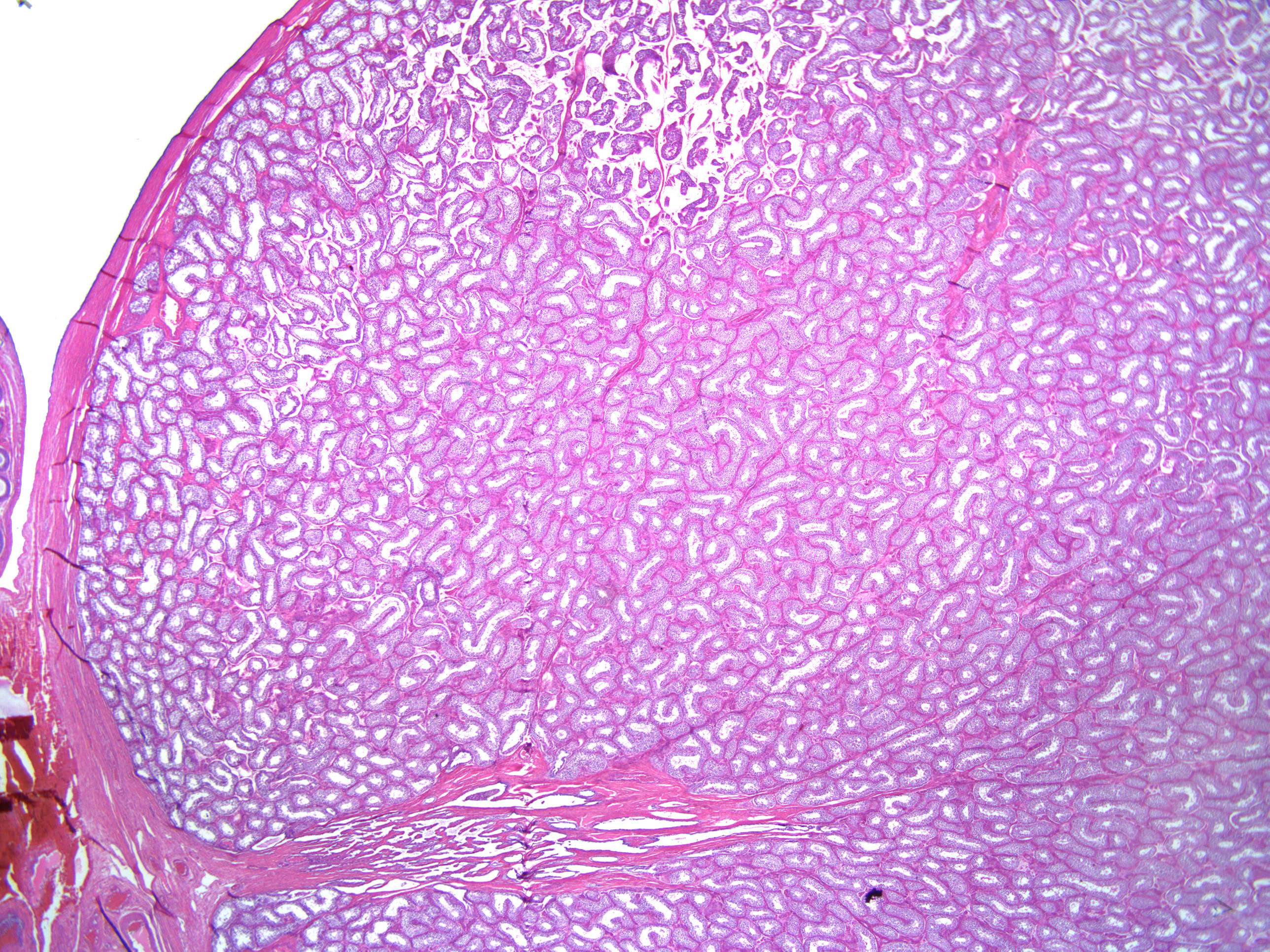
This is a higher magnification of the testis. The tunica vaginalis covers the tunica albuginea but cannot be discerned in this image. The tunica vaginalis testis is a thin layer of mesothelium (peritoneal epithelium). It is a rest of the peritoneal process (processus vaginalis) that extended into the scrotum in embryonic life.