
Testis from an adult
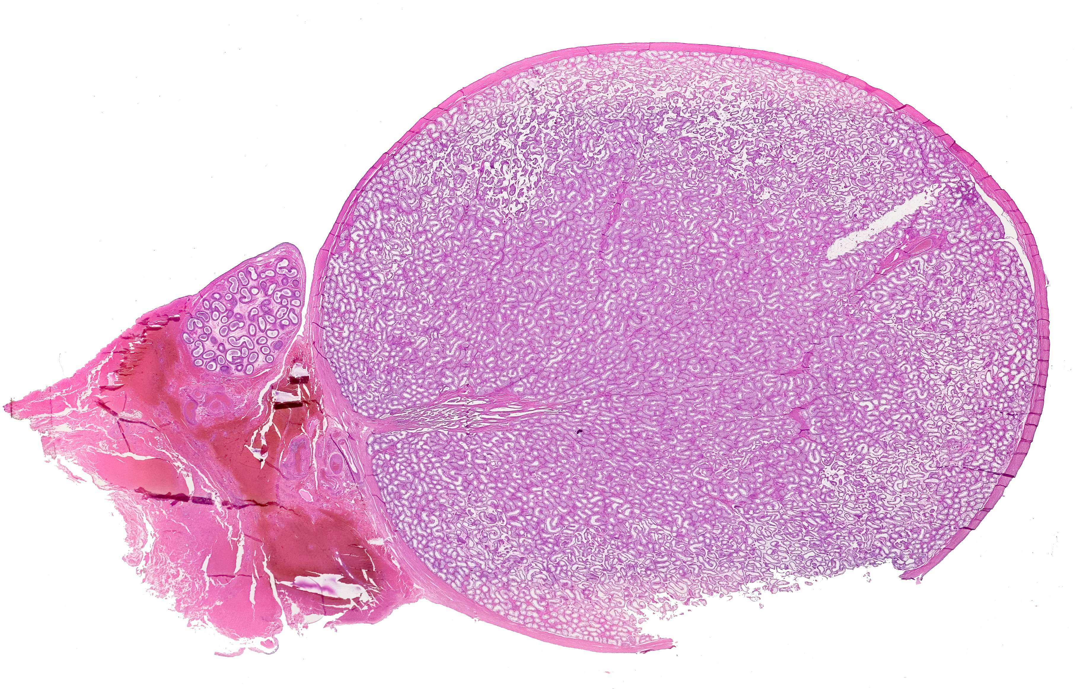
This is a low power photomicrograph of a testicle. To the left you can see a smaller structure which is the epidydimis. The testicle is covered by a thick and indistensible tunica albuginea, which consists of dense connective tissue and smooth muscle cells. It appears as a reddish band in the periphery of the testis.