

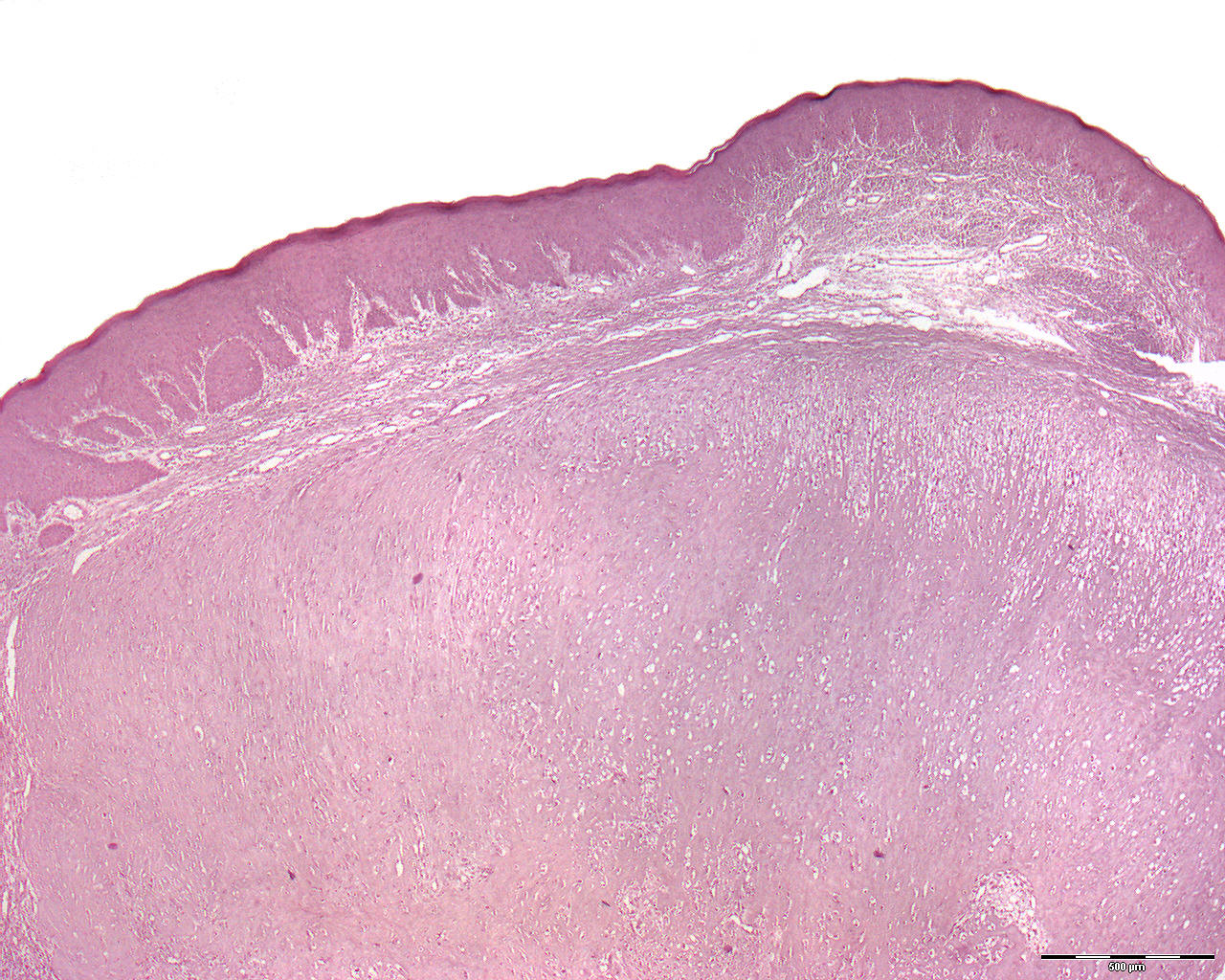
Osteosarcoma, chondroblastic type (40X)
The tissue is from the same patient as the other osteosarcoma image and originates from a resection of the left mandibular half during which the condyle was also removed.
Clinical diagnosis: Tumor mandibulae dext.
Microscopic examination: One finds pieces of mucosa with a convex surface covered by regular, well-defined multi-layered squamous epithelium with some presence of mononuclear inflammatory cells in the underlying, loose connective tissue. Below the epithelium, the tumor tissue is seen. It's quite fibrogenic with angular, oblong, coil-shaped, round and oval nuclei of varying sizes and coloration. Towards the resection margin deep part of the tissue, one might see a transition to malignant osteoid tissue (intramembranous ossification). One also finds cartilaginous areas with some variation in the size, shape and coloring of the cells.
Comment: Detection of malignant bone of the intramembranous type is a prerequisite for the diagnosis of osteosarcoma. If significant amounts of malignant cartilage are also present, the picture is called chondroblastic osteosarcoma.
Further: An en bloc (French, in block) resection of the mandible was carried out. Since then, the tumor has recurred numerous times, and it also arose contralaterally without affecting the midline. Today, the patient has had a total mandibulectomy.