


Intramembranous ossification (40X)
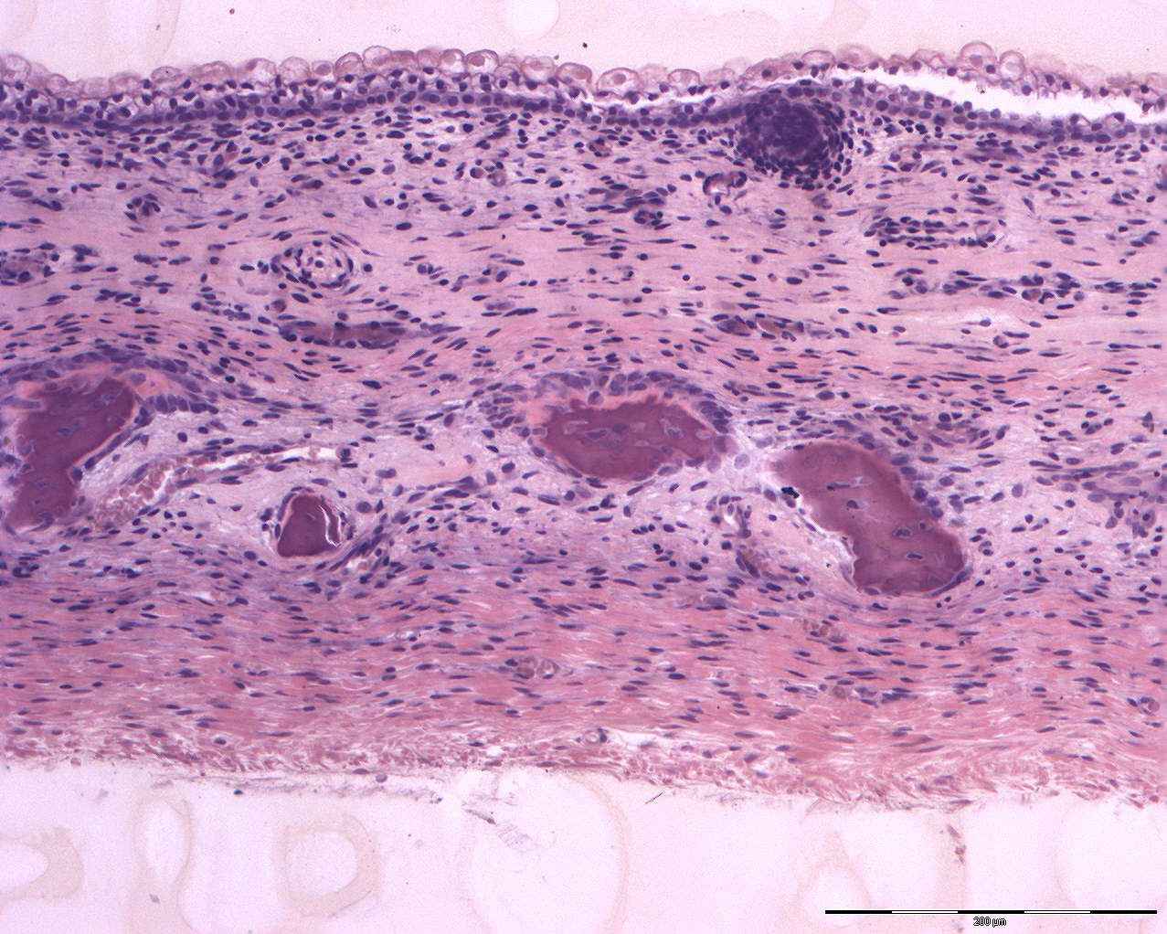
This section shows intramembranous ossification in the cranium.
The upper surface is covered by epithelium and just below the surface, an epithelial invagination is seen. This invagination is the preliminary stage of a hair follicle.
In the middle of the image, you can see "islands of bone" as larger chunks of purple matter. Osteocytes are embedded in these "islands". The osteocytes reside inside small cavities in the bone called lacunae.