

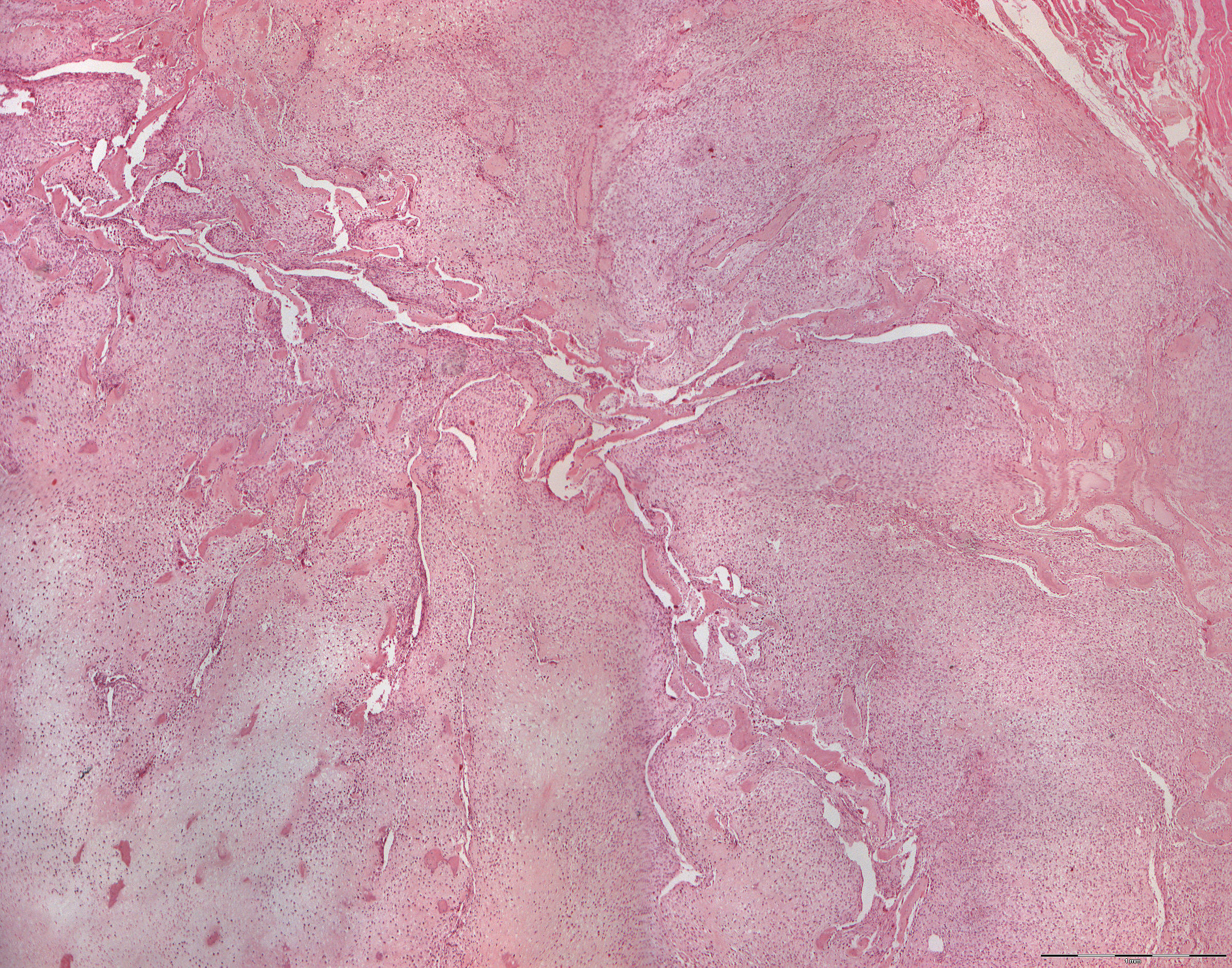
Osteosarcoma histology (40X)
Clinical information: A 23-year-old woman presented her with a 4 month history of swelling adjacent to teeth 45 to 47. Both 47 and 46 were extracted and a biopsy of the bone was taken (section no. 193).
Microscopic examination: Tumor masses are found which are peripherally limited by a narrow rim of connective tissue. Beyond this, you will find some skeletal muscle. Cortical bone tissue is not present. In front of the connective tissue barrier, a highly cell-rich, irregular, chondroid tissue and polymorphic cells are seen. You will find a number of bone trabeculae which, in any case, clearly originates from cartilage in some places. More centrally, areas with feww cells are found which stain weakly eosinophilic and where the chondrocytes are very swollen. Light, necrotic areas and some bleeding are seen. Particularly towards the necrotic part, residual trabeculae of sclerotic bone tissue (not tumor bone) with signs of resorption are detected.