

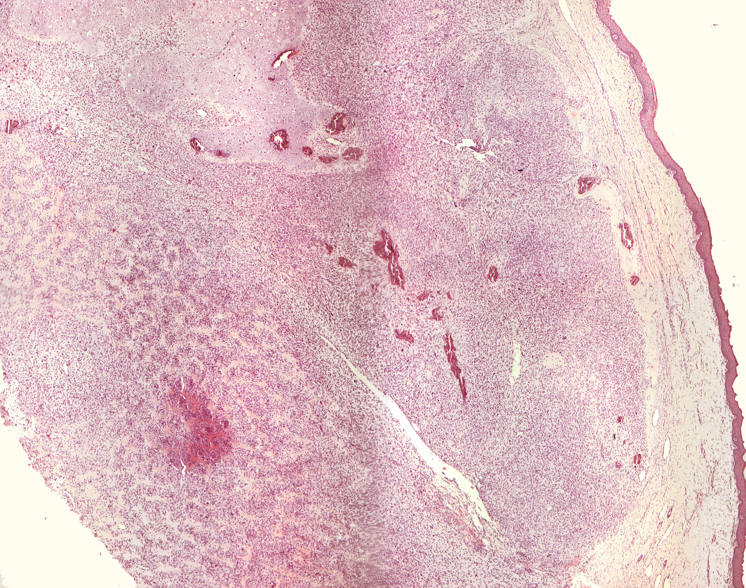
Osteosarcoma, chondroblastic type (40X)
Clinical information: In a 29-year-old man, there was a partially ulcerated swelling in buccal region of tooth 23 that had been there for about of half a year's duration. All the teeth on the left side of the upper jaw reacted negatively to the vitality test, while the teeth on the right side were vital. A biopsy was taken from the swelling.
Microscopic examination: One finds mucosal tissue samples covered by regular multilayered squamous epithelium with an underlying narrow, inconspicuous connective tissue zone. Slightly deeper, tumor tissue is seen with a certain fibrogenesis and made up of densely packed, undifferentiated, mesenchymal and partly fibroblast-like cells with pronounced nuclear polymorphism and the presence of multinuclear tumor giant cells. Even deeper in the tissue, traces of intramembranous bone with some mineralization (basophilic reaction) are seen. Polymorphic cells and malignant osteoblasts surround this bone tissue. There are also larger areas made up of hyaline cartilage with some mineralization and considerable variation in the chondrocytes' size, shape and coloration. The malignant connective tissue cells are sometimes seen in very close relation to small veins and in some places probably intravascularly (haematogenous spread).
Comment: Occurrence of malignant connective tissue of intramembranous bone is a prerequisite for the diagnosis of osteosarcoma. This can also, as here, contain significant amounts of fibroblast-containing and undifferentiated mesenchymal tissue and cartilage tissue. Depending on the predominant tissue type, osteosarcomas are divided into fibroblastic, osteoblastic and chondroblastic types. Long tubular bones are the predilection site for osteosarcomas, which are preferably seen in young boys/younger men. Osteosarcomas in the jaws tend to occur in slightly older patients, as in this case, and the prognosis is better than for osteosarcomas in long tubular bones, especially for mandibular tumours.
X-ray examination after the histological diagnosis had been made showed extensive tumor masses in his left maxillary sinus and small perforations of the bottom of the orbita. The patient underwent preoperative immunosuppressive therapy to reduce tumor activity before hemimaxillectomy hoping to retain the bottom of the orbita.