

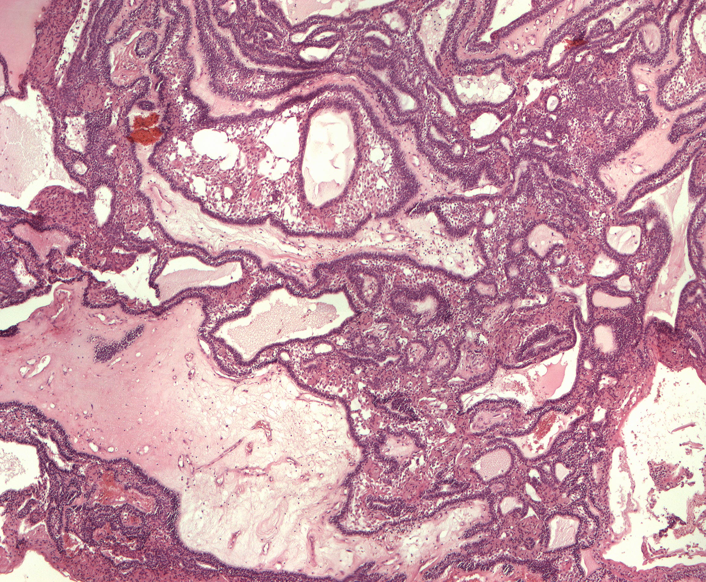
Plexiform ameloblastoma (40X)
Clinical information: A 77-year-old woman was operated on for a tumor in the left part of the mandible 40 and 32 years earlier. In recent years there have been tumor recurrences and currently she presents a fist-sized tumor in the left mandibular region. X-rays showed destruction of the mandible and only some thin bone lamellae surrounding it. During operative removal of the tumor, the proximal part of the mandible could not be located. The frontal part was resected (resecare, L = cut off again) all the wy to the symphysis. The tumor was multilocular on sectioning.
Clinical diagnosis: Tumor mandibulae sinistra. Ameloblastoma.
Microscopic examination: One can see pieces of tissue made up of epithelial tumor tissue which mostly grow in a net-like pattern (plexiform). There are also islands (follicles) of varying sizes and sometimes with significant central, cystic decay. Peripherally, low, cylindrical cells resembling ameloblasts or preameloblasts are found in the tumor tissue. Centrally, densely packed, polygonal cells are mostly seen, but also areas of resemblance to the star-shaped reticulum of the enamel organ. Larger and smaller accumulations of large, eosinophilic, granular cells with a relatively small, dark nucleus (granular cells) are found in places in the epithelium. Some degenerative changes are seen in the surrounding stroma and partly pronounced circulatory disturbances due to how the plexiform tumor proliferations compromise the blood supply. Electron microscopically and histochemically, the granules resemble lysosomes.