


Early stage of a hair follicle (600X)
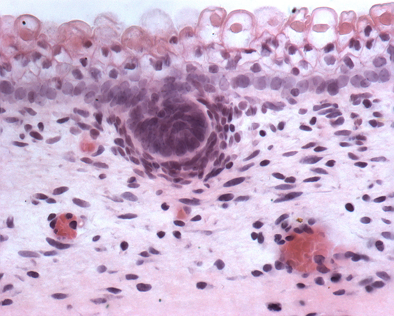
The surface is covered by epithelial cells. The nucleus can easily be seen inside the cells. Jjust below the surface an epithelial invagination is seen. This invagination is the preliminary stage of a hair follicle. Two blood vessels filled with erythrocytes are also seen.