


Cementum, periodontal ligament and bone (100X)
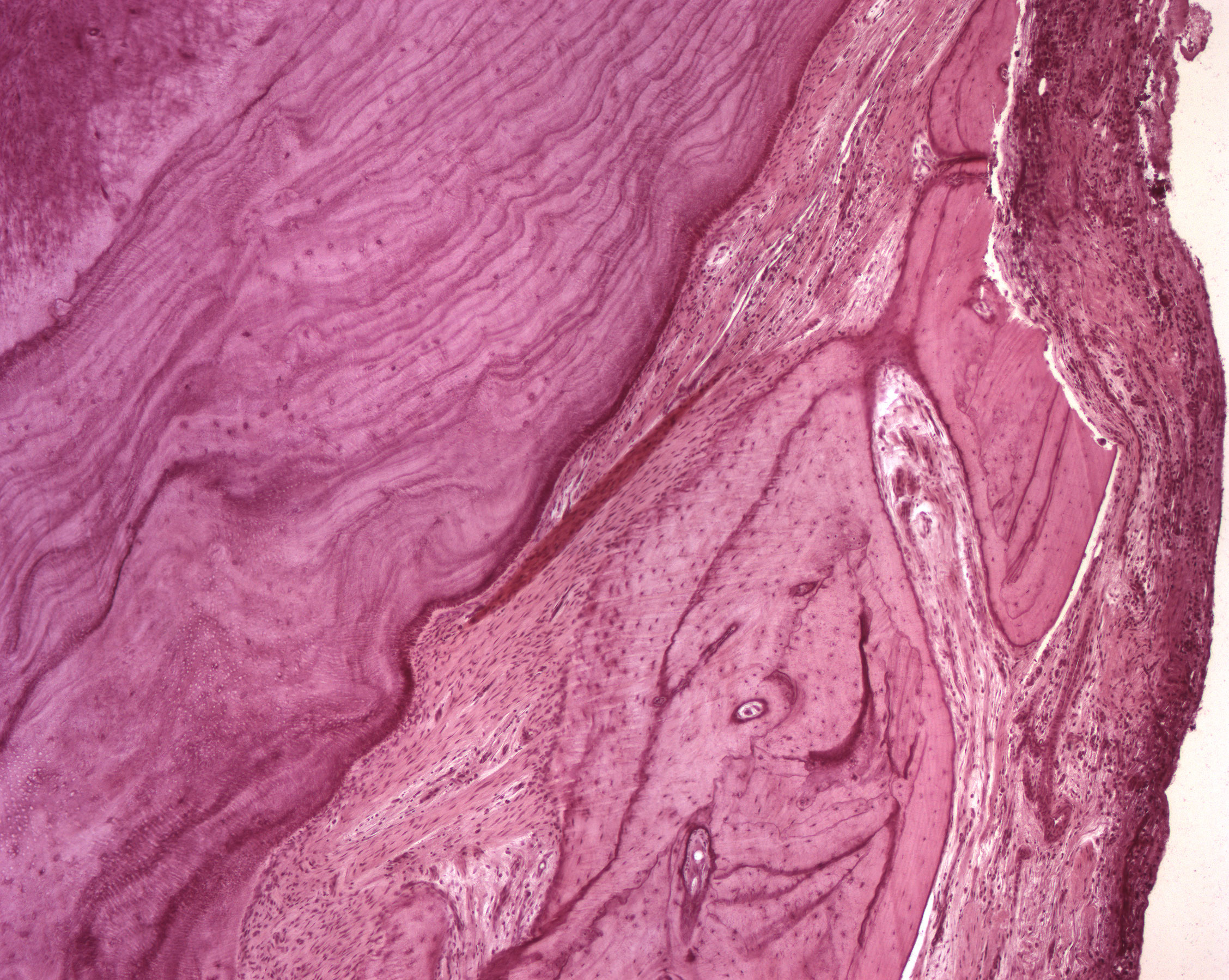
Here's a magnification of the cementum. As mentioned, the cementum is mostly of the cellular type and cementocytes are scattered throughout the cementum. Numerous incremental lines can be seen and illustrates the "rhythmical" deposition of cementum. The periodontal ligament (PDL) is seen more peripherally to the cementum followed by the alveolar bone. Osteocytes reside in the bone.