


Apex of a root with cementum (40X)
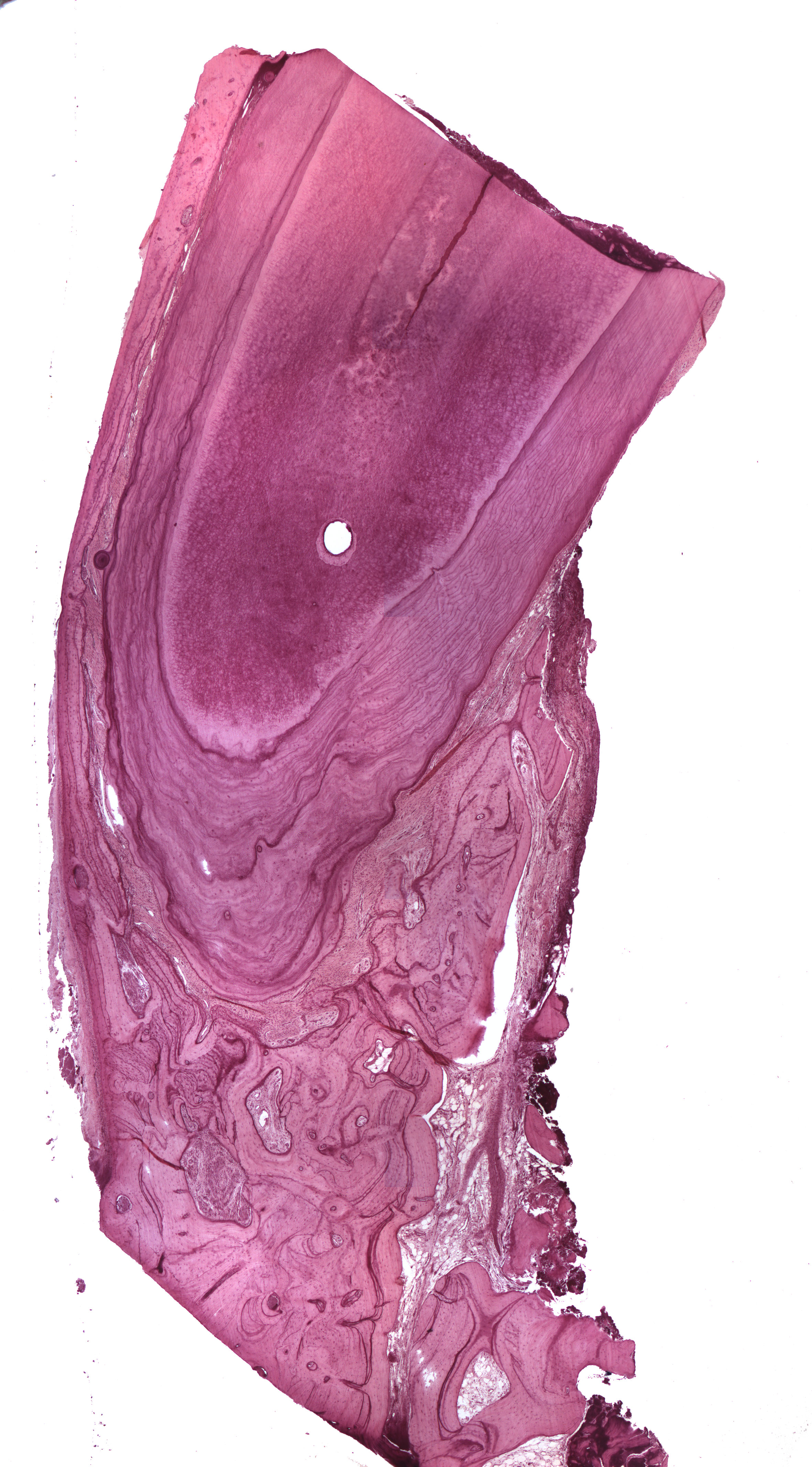
This is a longitudinal section of the apical part of a root and the image is well suited for taking a closer look at the cementum. The apical supportive tissues are also seen, but no apical foramen. You may already have noticed the thick layer of cementum covering the apical part of the dentine. You may also have made the thought that this tooth once belonged to an old individual. If that's right, you can pat yourself on the back because it's true! This is because the cementum is thick and mostly of the cellular type. There are many incremental lines which illustrate the "rhythmical" deposition of cementum through many years.
Use the buttons below the image to see the rest of the pictures for this section (i.e. the apex).