


Cornea with anetrior corneal epithelium (400X)
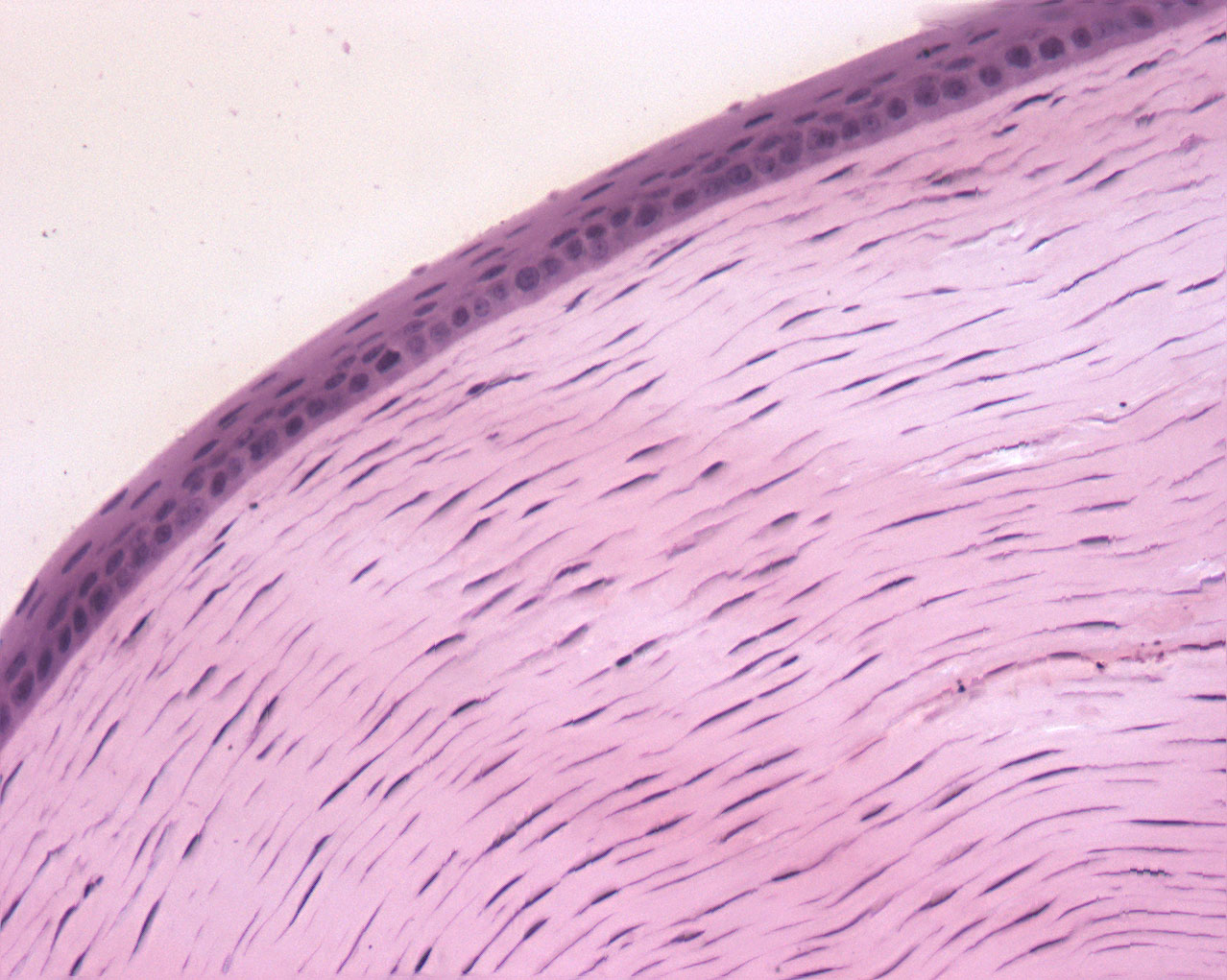
The anterior surface of the cornea is covered by squamous apithelium (lat: epithelium anterius corneæ); the posterior surface is covered by quboidal cells (not visible in this image). Both of these epithelial membranes are separated by the interior of the cornea by thin membranes structureless membrane.
The collegenous lamellae of the cornea can easily be seen because of the adjacent fibroblasts. In a healthy eye, all light needs to travel through all these layers, hence the cornea is totally transparent. Any illnesses in the cornea might affect your vision.
Notice that there are no blood vessels in the cornea.