


Cornea and eyelid (400X)
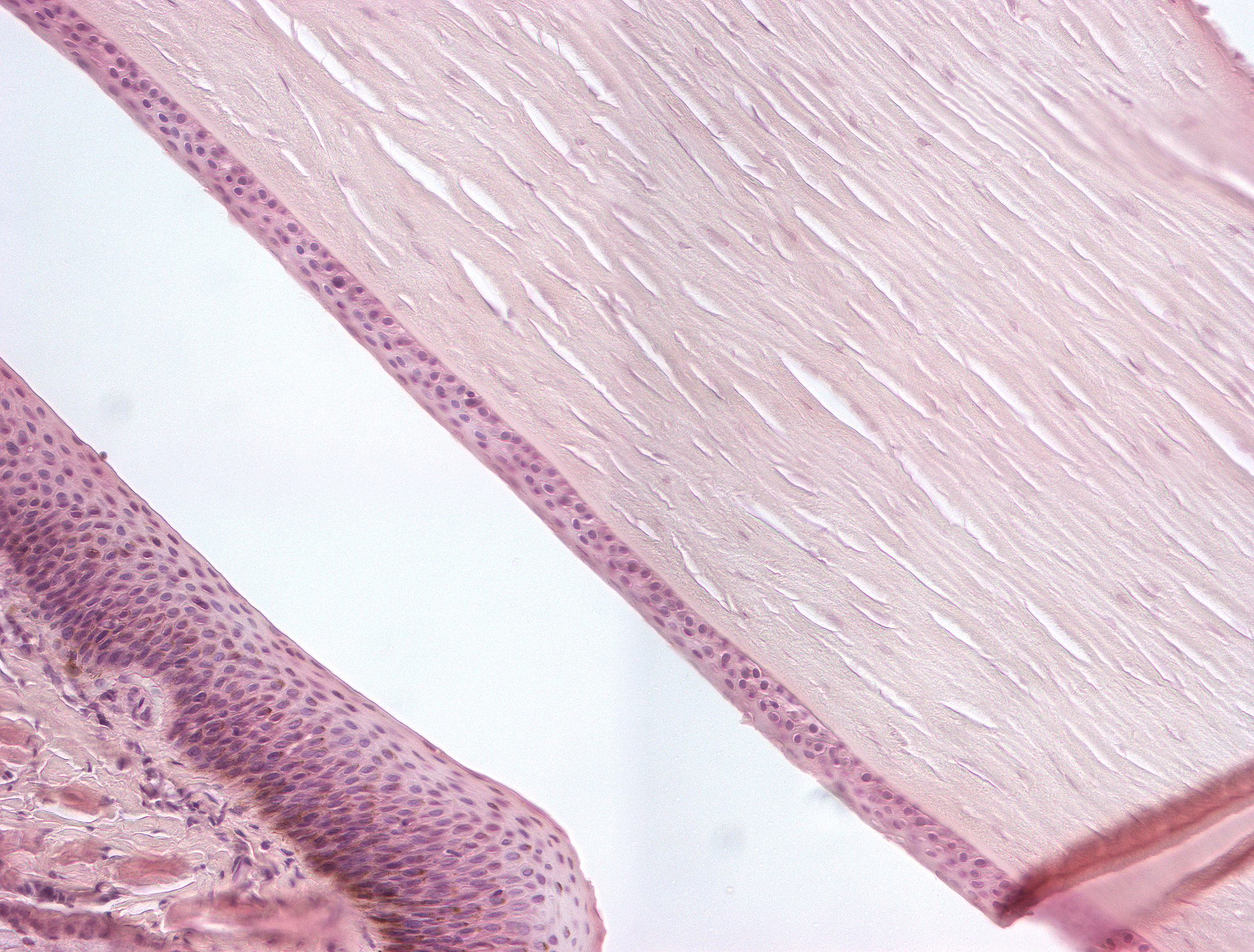
At higher magnification, you can actually see the epithelium covering the cornea. The outer surface is covered by stratified squamous epithelium (lat: epithelium anterius corneae). The posterior surface of the cornea is also covered by epithelium, but it is not as thick (lat: epithelium posterius corneae). As you can see, the cornea has no blood supply and relies solely on oxygen from the air that surrounds the cornea naturally. The structure of the inner layers of the cornea is also well depicted with its lamella of collagen fibrils and fibroblasts.