


Sublingual gland (600X)
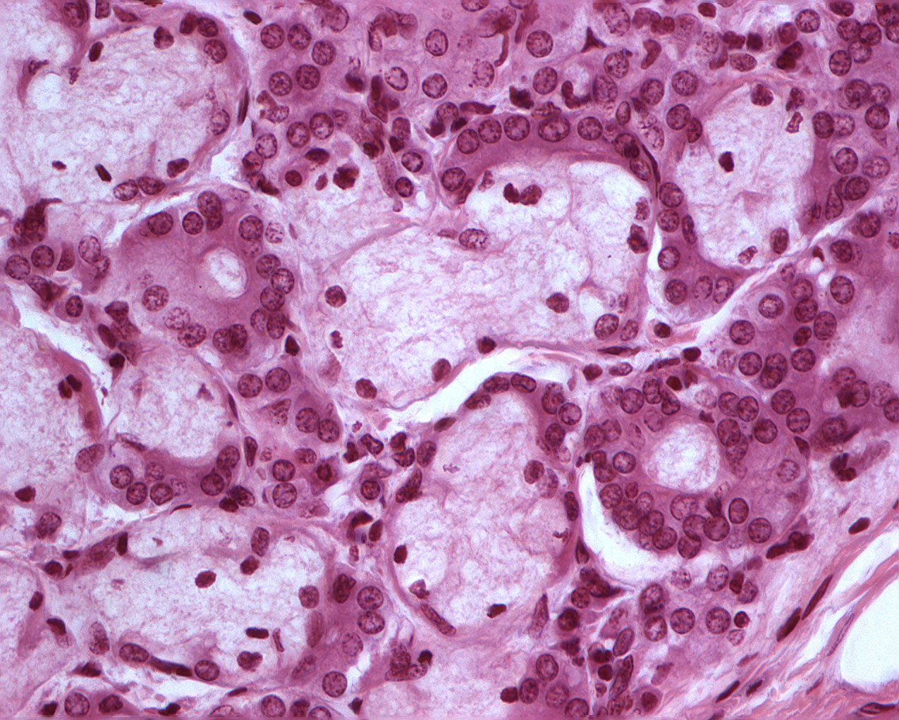
This image shows details of the acini and the excretory duct. There are mucous cells surrounded by a semi lunar looking cluster of serous cells (serous demilunes).
The lumen of the excretory ducts are easy to spot. That goes for the chromatin in the nuclei of cells of the excretory ducts and the serous cells too.