


Sublingual gland (400X)
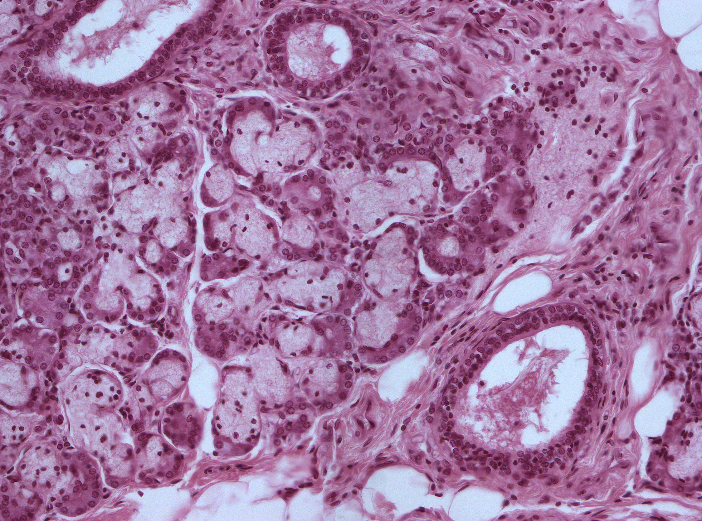
As you can see, the mucous acini dominate. Basally to the cells of the mucous acini, one can see semilunar serous cells (darker cells making upm the serous demilunes). There are also myoepithelial cells between the acini, but they are hard to spot and are hard to differentiate from fibroblasts in this image.