


Sublingual gland (100X)
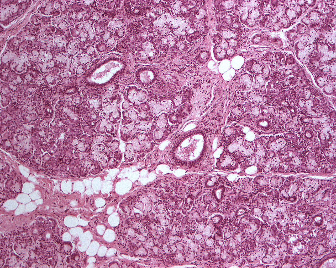
The mucous acini dominate. Most mucous acini are capped with serous demilunes
You won't see as many intercalated ducts in the glandula sublingualis as in the parotid and submandibular glands because the intercalated ducts are very short in this gland.
Within the septa of connective tissue one can find large excretory ducts, arteries and veins. One can also spot adipose tissue (large white cells).