

Muscle spindle in a transversal section of skeletal muscle (600X)
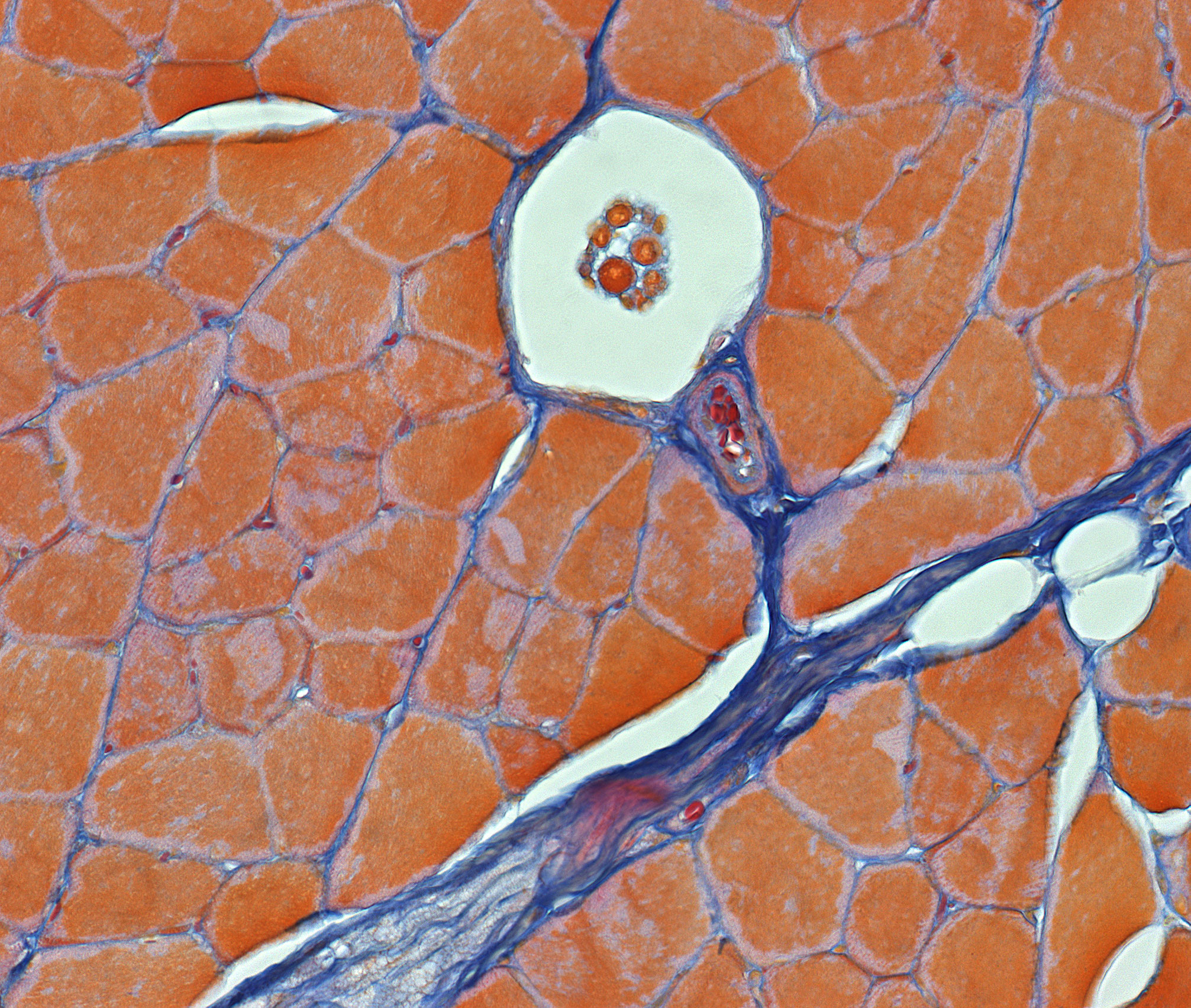
This image shows details from a transversal section of a skeletal muscle. In between the larger muscle fibers and contained in the perimysium, you can find muscle spindles. Their main function is to detect changes in the length or stretch of the surrounding muscle fibers. The muscle spindles are for one thing made up of small muscle fibers called intrafusal fibers. These fibers are both innervated by afferent- and efferent neurons (see your textbook of physiology for further details on how this great organ works). The efferent innervation is provided by the γ-motoneurons (extrafusal fibers (normal skeletal muscle)) are innervated by α-motoneurons). The nerve fibers are hard to see in this section.
The muscle spindles belong to the proprioceptive branch of the senses.