
Pons histology (12.5X)
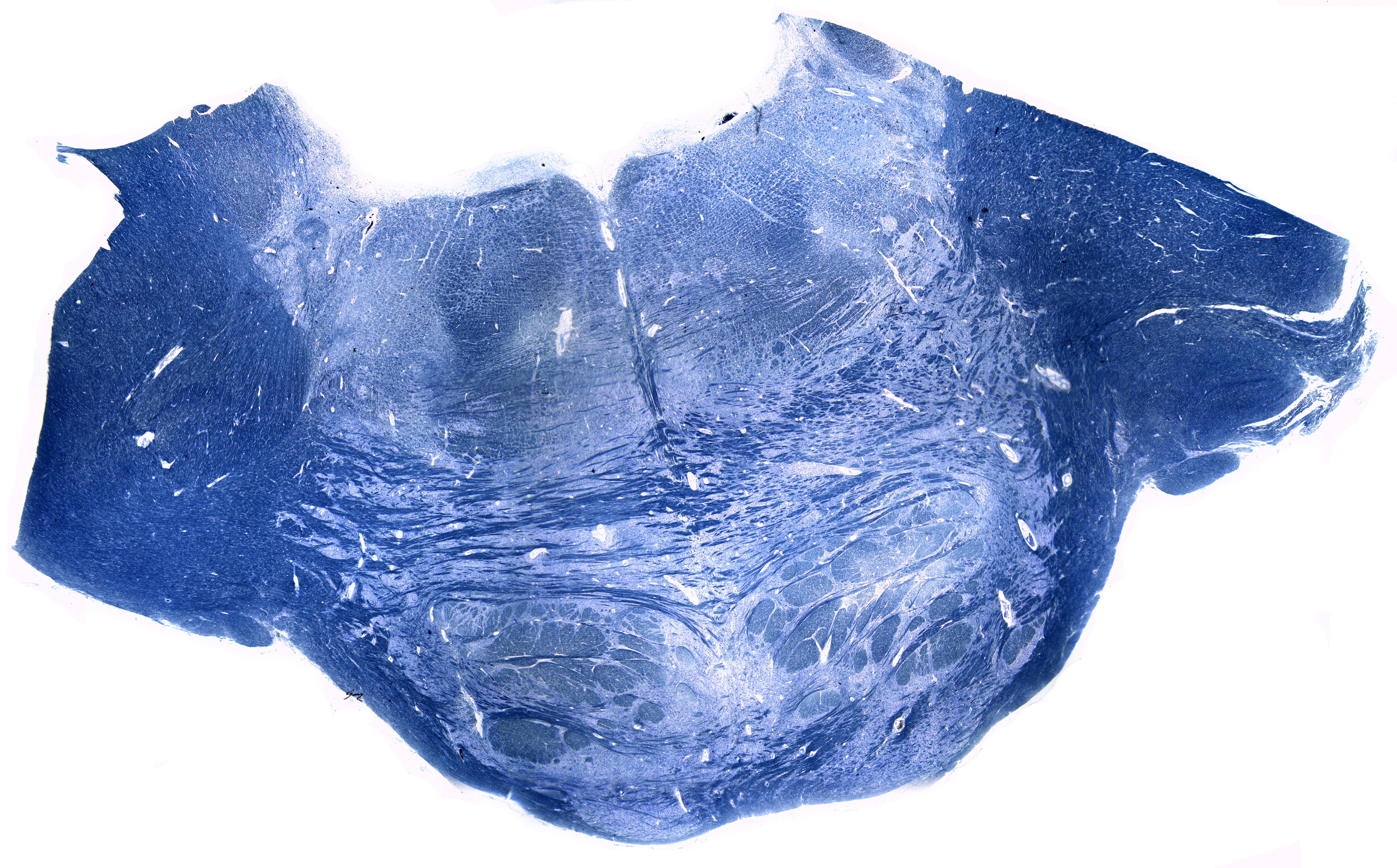
The image shows a transverse section through the middle part of the pons. It is stained in such a way that the myelin is coloured blue. The neurons themselves are not stained in this section.The pons contains a lot of important structures that you've probably heard of. At first glance it might be overwhelming to try to distinguish them from eachother. But try to point out the following structures:
- Reticular formation
- Superior cerebellar peduncle
- Middle cerebellar peduncle
- Medial lemniscus
- Pyramidal tracts
- Trigeminal motor nucleus
- Trigeminal sensory nucleus
- Medial longitudinal fasciculus
- Pontine nuclei
The pyramidal tracts are aggregations of efferent nerve fibers from the upper motor neurons that travel from the cerebral cortex and terminate either in the spinal cord and are involved in the control of motor functions of the body.