
The medulla oblongata - overview (12.5X)
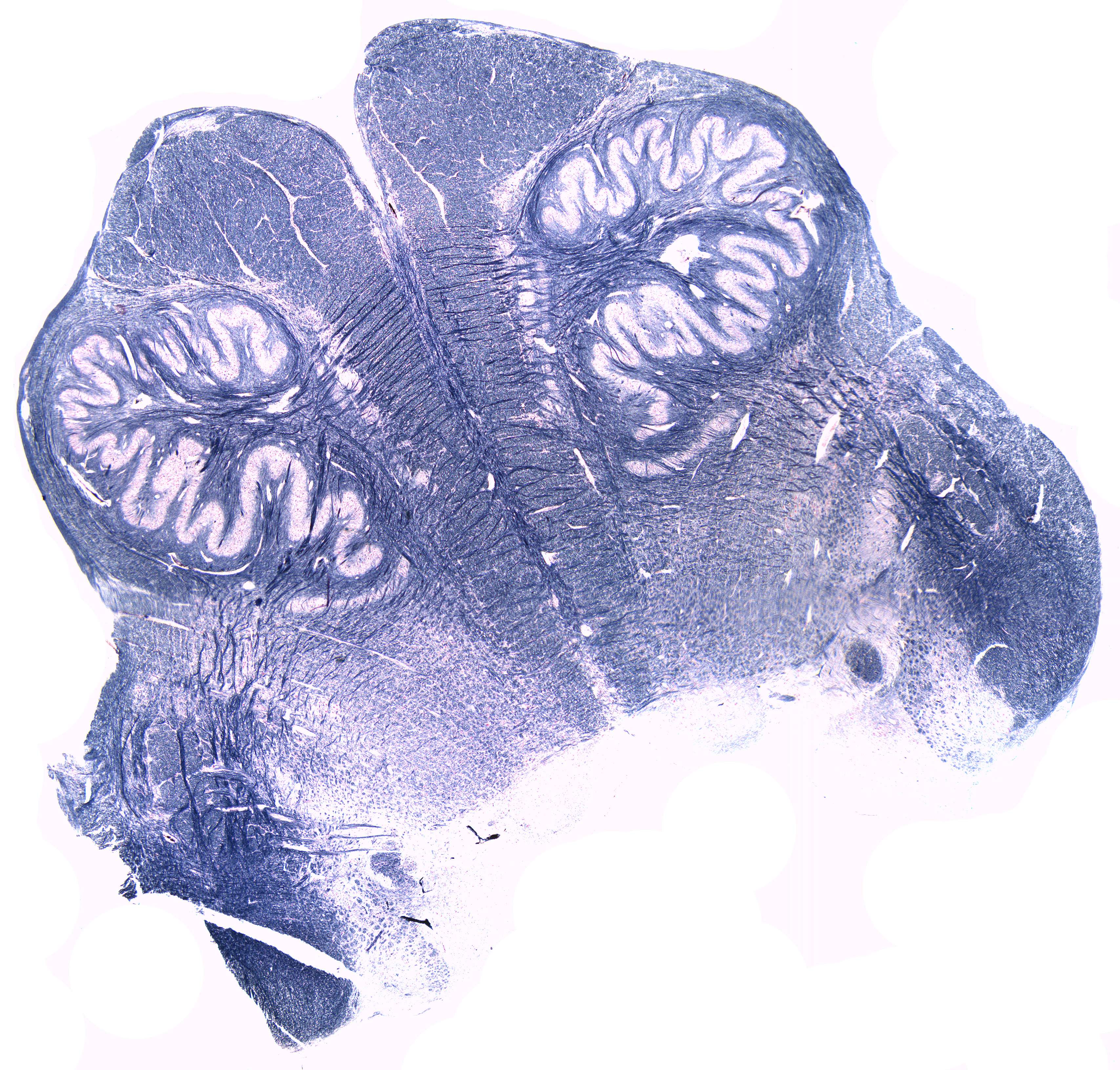
This is a cross-section through the upper part of the medulla oblongata stained in such a way that the myelin obtains a blue colour while the nerve cells have a reddish look.
The sensory trigeminal nerve nuclei are the largest of the cranial nerve nuclei, and extend through the whole of the midbrain, pons and medulla, and into the high cervical spinal cord.
The spinothalamic tract is a part of the anterolateral system or the ventrolateral system, a sensory pathway to the thalamus.
The medial lemniscus is a large ascending bundle of heavily myelinated axons that decussate (cross) in the medulla oblongata. The medial lemniscus is formed by the crossings of the internal arcuate fibers. The medial lemniscus is part of the dorsal column–medial lemniscus pathway, which ascends from the skin to the thalamus, which is important for somatosensation from the skin and joints. Therefore, lesion of the medial lemnisci causes an impairment of vibratory and touch-pressure sense.
The inferior olivary nucleus is known to coordinate signals from the spinal cord to the cerebellum to regulate motor coordination and learning. As you can see, the inferior olivary nucleus has a distinct laminar structure. These lamina house the cell bodies of the olivocerebellar fibers. These neurons are the major input source for the cerebellum. Their axons are referred to as climbing fibers.
The medullary pyramids are paired white matter structures of the brainstem's medulla oblongata. The two pyramids contain the motor fibers that pass from the brain to the medulla oblongata and spinal cord.