
Jejunum
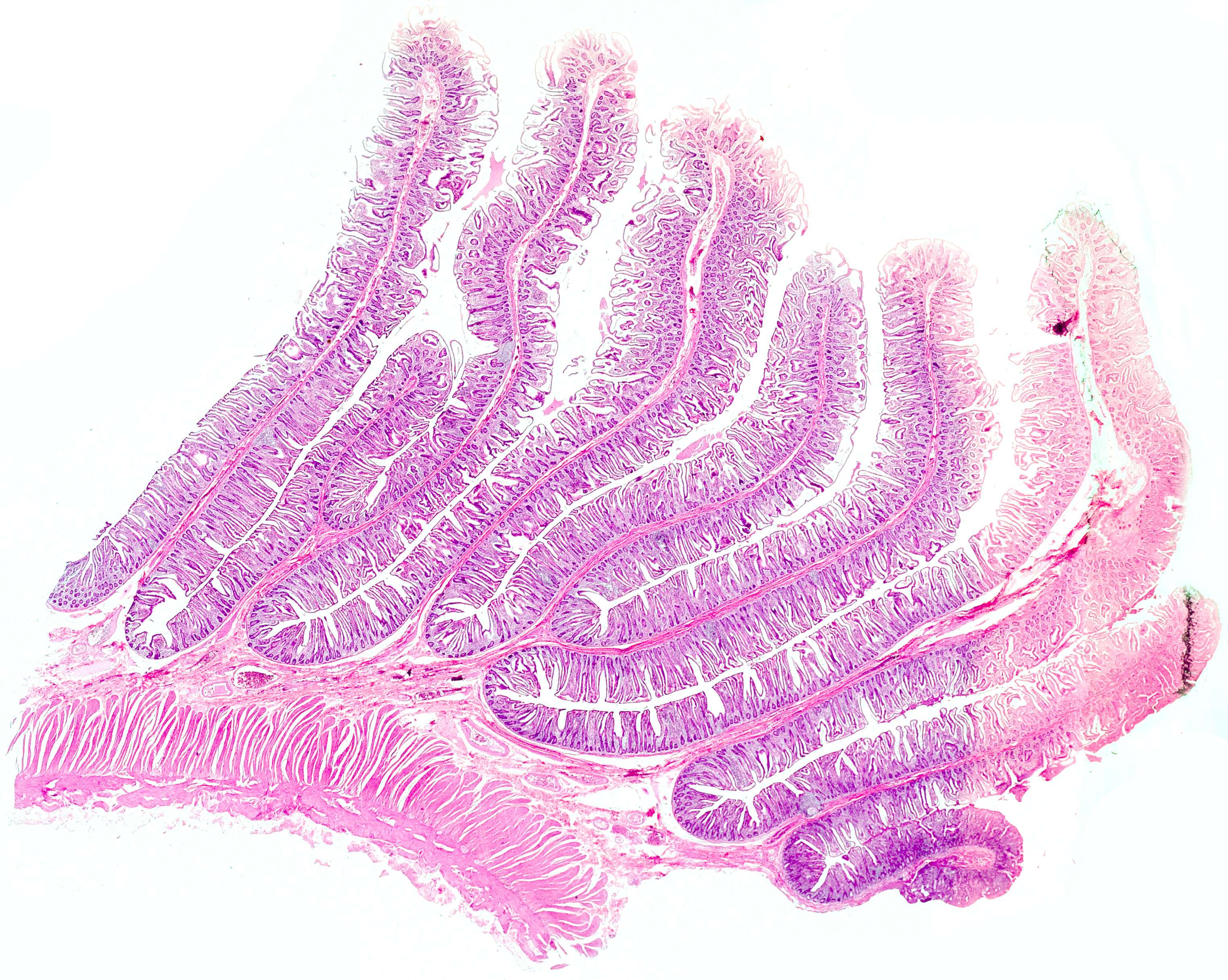
Macroscopically, the tissue is very folded. Microscopically, connective tissue features are seen edged with mucosa on both sides. These structures are called plicae circulares and help to increase the surface of the intestinal mucosa. Note that the incision is cut across the plica, and thus in the longitudinal direction of the intestine.