


The temporomandibular joint (20X)
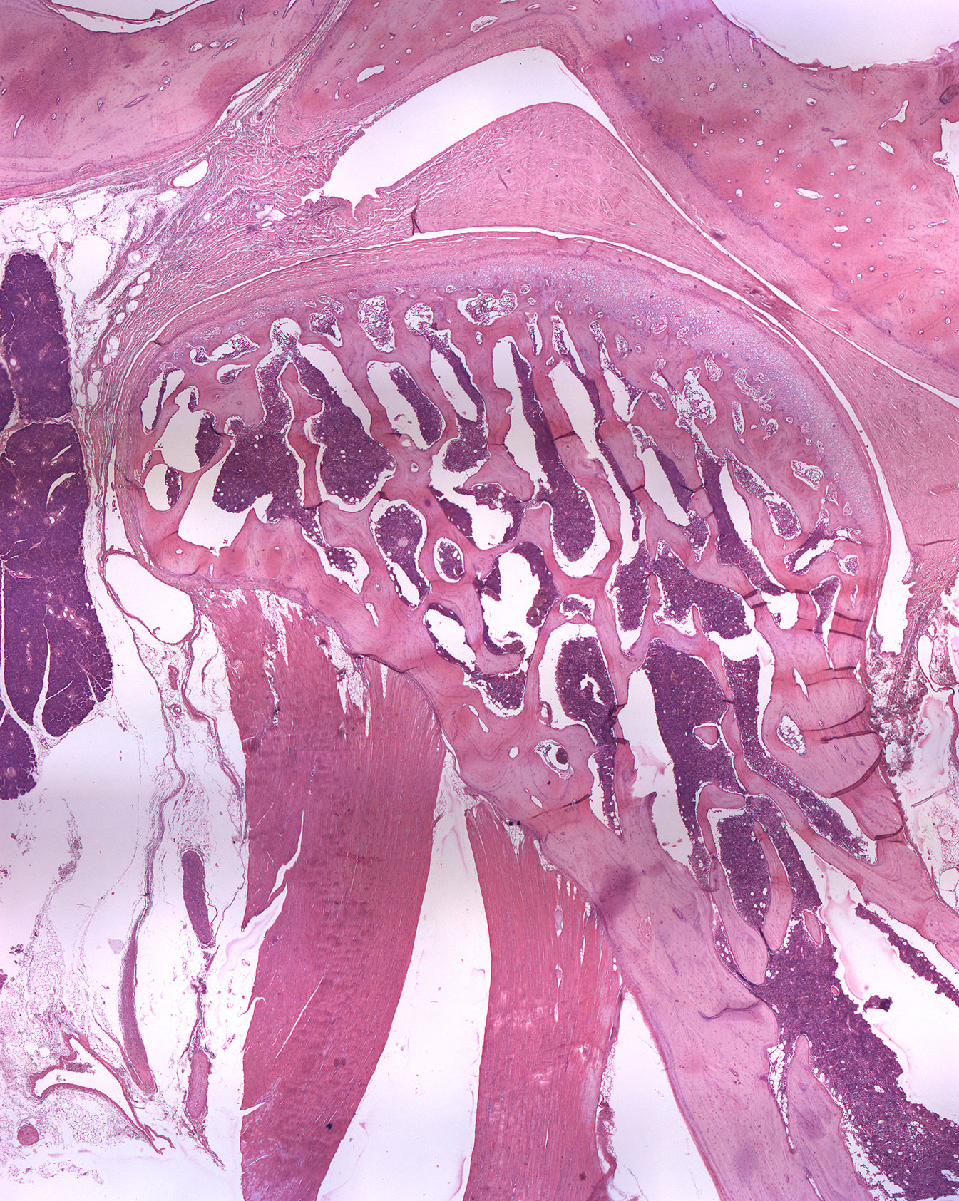
This is an H+E stained saggital section through the temporomandibular joint (TMJ) of a monkey. It is evident that the condylus is still proliferating. You can see the temporal bone with its mandibular fossa covered by a thick, fibrous tissue containing fibroblasts. The bone of the condyle is filled with bone marrow. Both muscle- and glandular tissue is seen in this section.