


Serous acini and striated duct (600X)
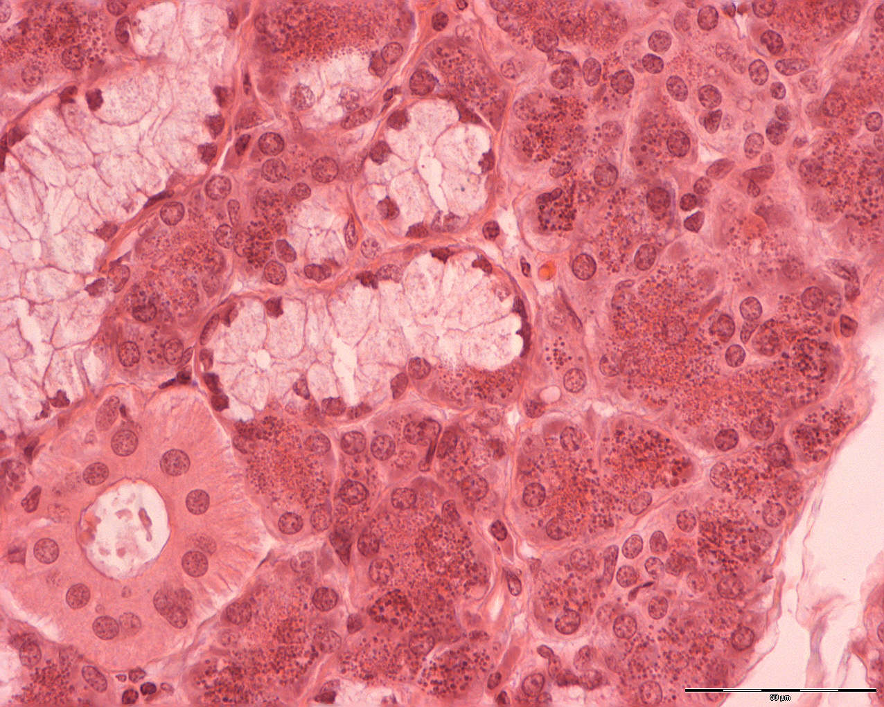
At higher magnification, you can clearly see the serous acini. The nucleus is round and situated basally in the cells of the acinus. These acini stain darker than their counterpart, the mucous acini. They are larger than the serous acini and their cells stain less. The nuclei of the mucous cells are flattened and situated at the base of the cell membrane. You can also see a striated duct, getting its name from of its striation (or stripes).
The mixed acini are normally mixed mucous acini surrounded by a group of serous cells. In the microscope it looks like a crescent-shaped dark moon (serous demilune).