
Epididymis
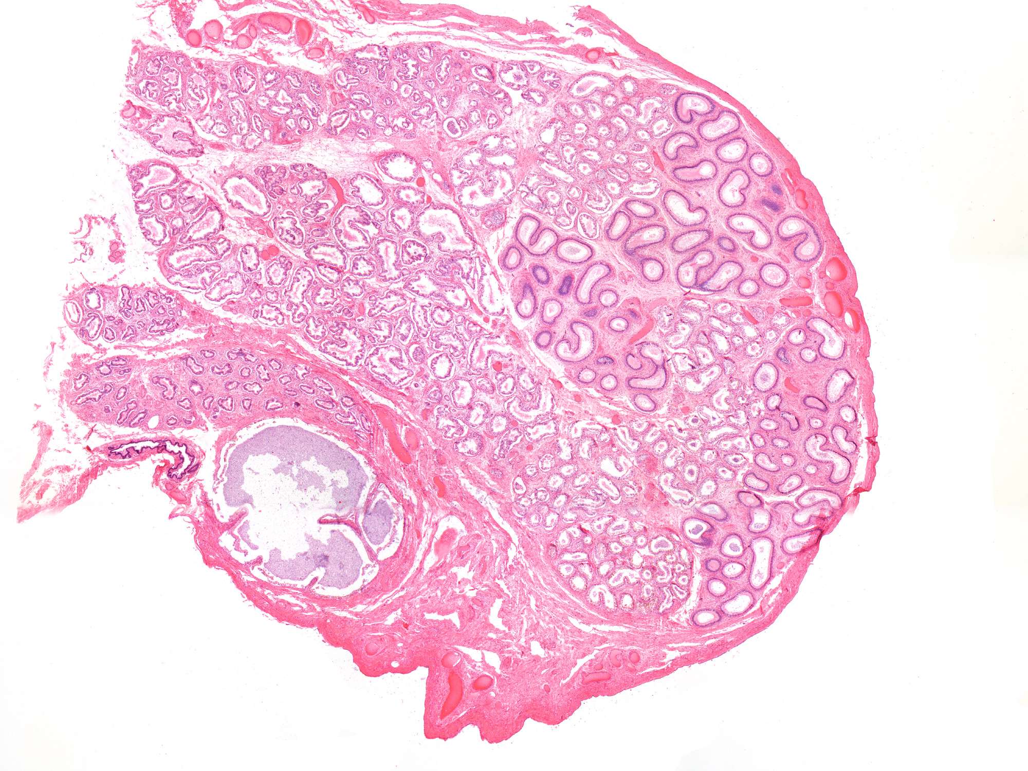
This is a low-power photomicrograph of a section through the epididymis. Many tubular structures are visible, and you might see that there are two types of tubules. The epididymis is covered by a tunica albuginea, appearing as a pink layer (note that it is thinner than the corresponding layer in the testis). Outermost is a very thin layer of tunica vaginalis (cannot be identified at this magnification).