

Tooth with apical granuloma and apical periodontitis
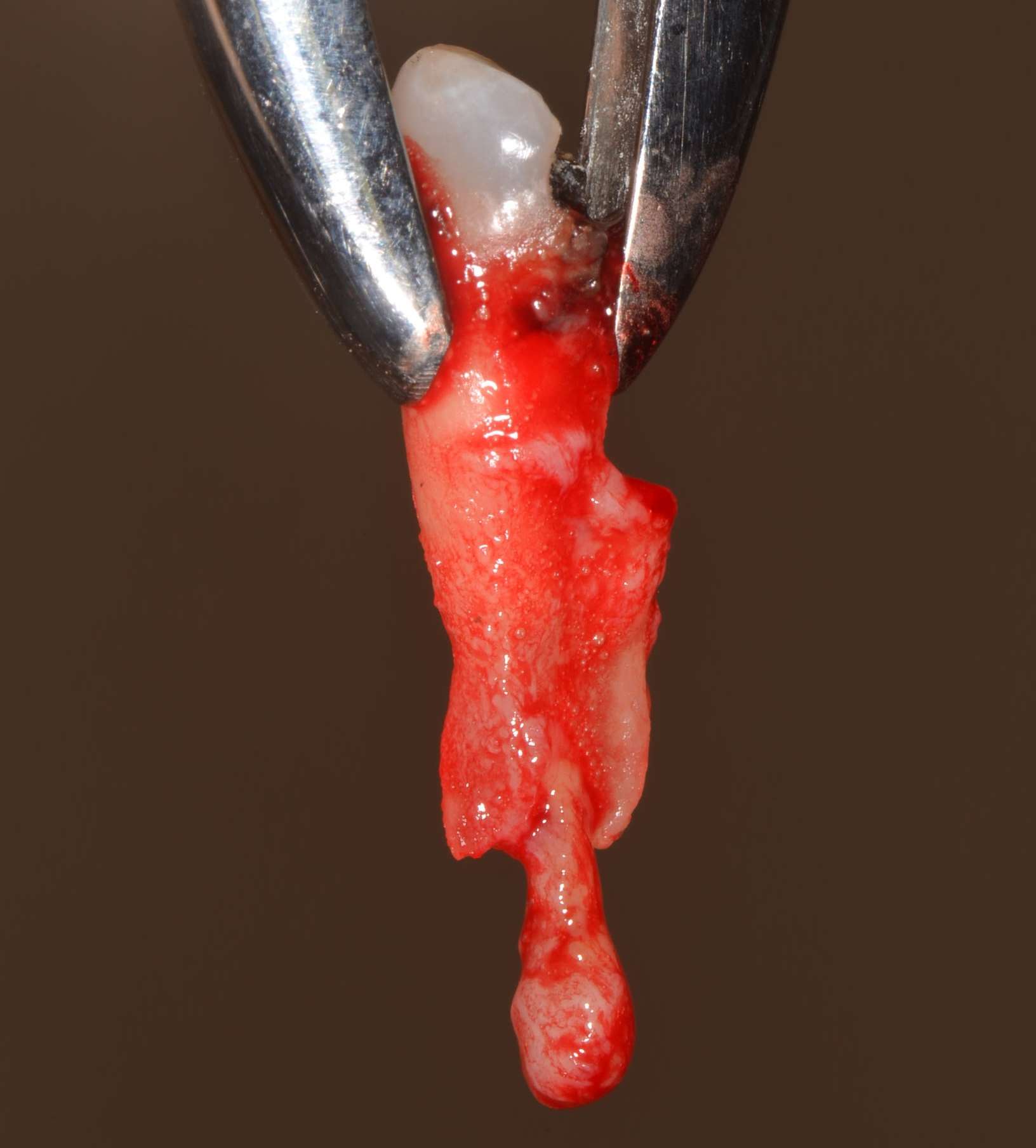
The image shows a freshly extracted premolar. This is a premolar from the upper jaw (either 14 or 24) as you can distinuish two thin gracile roots. A forceps used to remove the tooth fixates the tooth firmly at its cervix. Between the two parts of the working end of the pliers you can see the enamel in the crown of the tooth.
Apical to the root tip (apex) there is a granuloma. This tissue is largely made up of inflammatory cells (lymphocytes, macrophages and a good number of granulocytes). See also the microscopic image of an apical granuloma.