

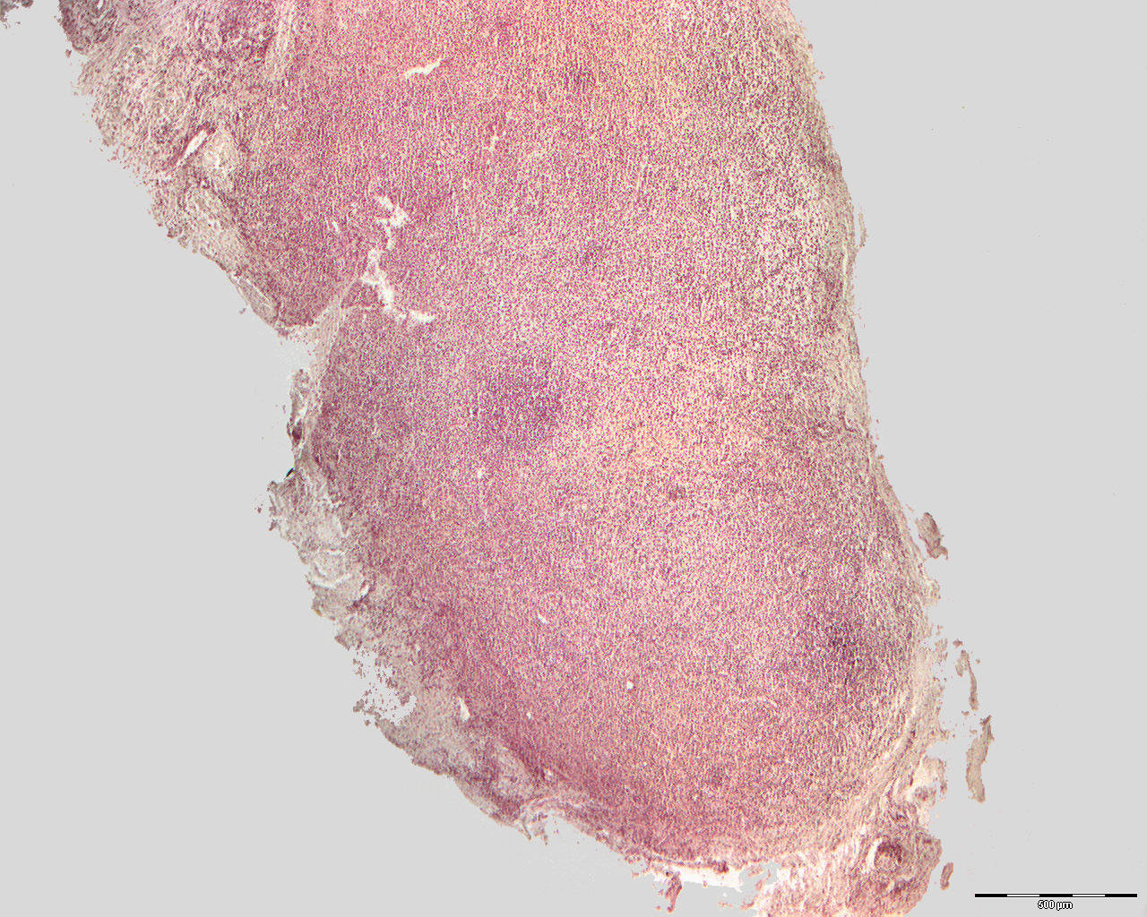
Granuloma from dental root and subacute inflammation (40X)
Clinical information: The patient was a 39-year-old man that have had some pain in the region of tooth 25. X-rays showed apical clearing. Soft tissue was removed. See clinical image of extracted premolar with apical granuloma.
Clinical diagnosis: Cystis apicalis 25.
Microscopic examination: You can see many pieces of soft tissue, some of which display a very irregular, small, central lumen filled with eosinophilic, herniated substance (coagulated proteins). Towards the lumen, a thick layer of granulation tissue is seen with an abundant presence of both mononuclear inflammatory cells, mostly lymphocytes, plasma cells and macrophages, as well as a good number of granulocytes. The tissue is relatively rich in small dilated veins. Peripherally, a connective tissue capsule is seen with individual beams of cancellous bone tissue.