
Corpus luteum
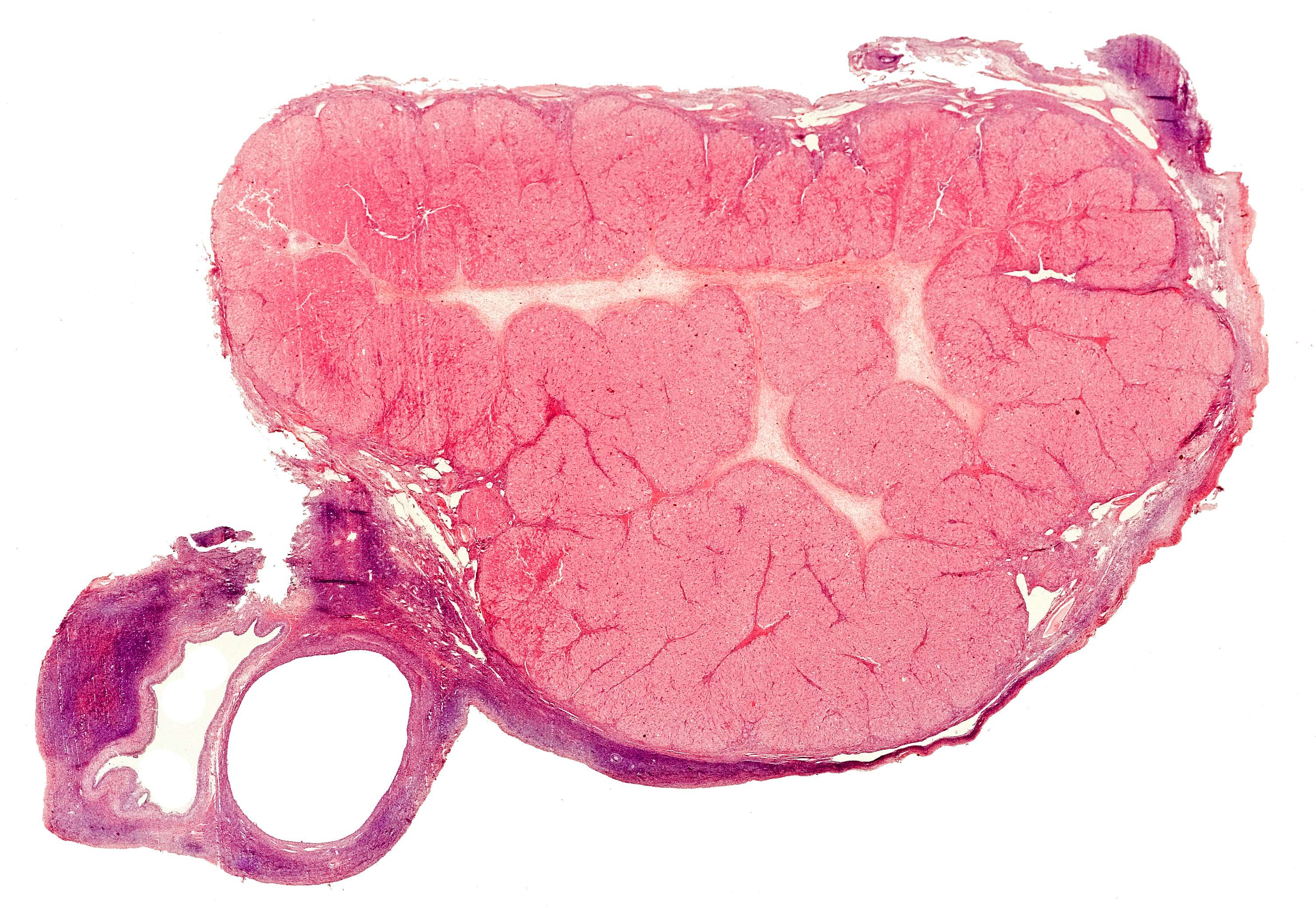
This is a section through a complete corpus luteum. The corpus luteum is made out of remnants of the follicle after ovulation has occurred.
You see a light colored area in the middle that consists of loose connective tissue and outside that, a convoluted reddish band (which consists of epithelial cells of the follicle, even if you cannot see cells at this magnification).
You may notice that the reddish band consists of somewhat differently colored areas; an outer thin, darker brim and a lighter inner main part. This represents the two kinds of endocrine cells in the corpus luteum (theca lutein cells and granulosa lutein cells, to be seen more clearly in the next photomicrographs). Ovarian tissue is seen down to the left and as a dark brim along the lower edge of the corpus luteum.