

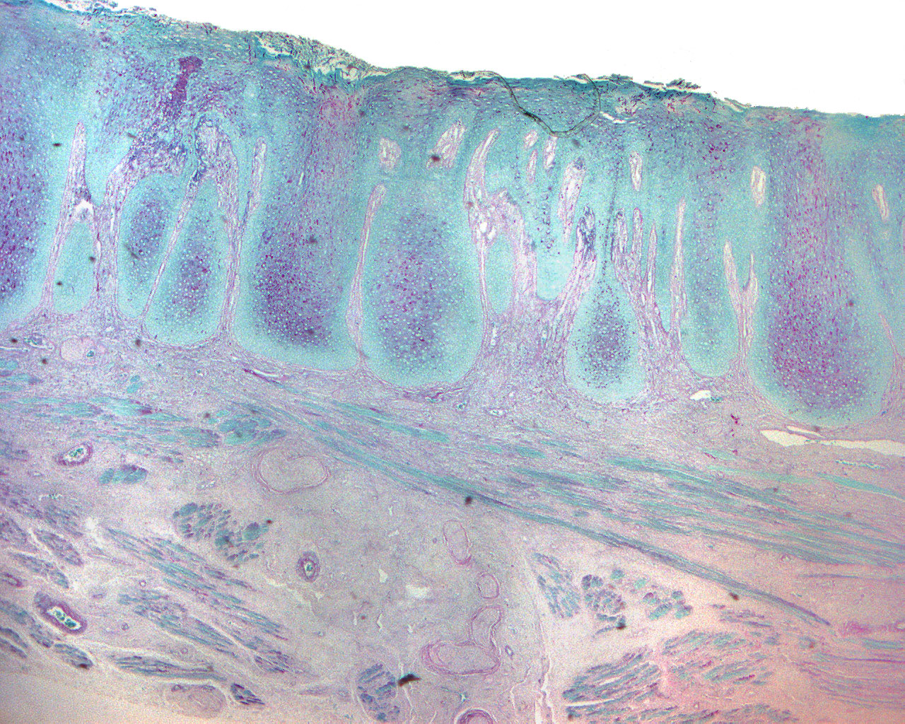
Fungal infected mucosa with squamous hyperplasia and moderate chronic inflammation (40X)
Clinical information: See this section for a H+E colored stain of the same tissue tample.
Microscopic examination: Special staining: (PAS/Light green) shows the local presence of numerous hyphae and pseudohyphae in the parakeratin layer. These are compatible with the pathogenic form of Candida species. The hyphae/pseudohyphae are often perpendicular to the epithelial surface. They grow intracellularly as parasites. Because they are so often located high in the stratum corneum, they are often lost with the surface layer during biopsy and laboratory procedures. If Candida infection is suspected, it is therefore essential to also take a microbiological sample. It is important to know that Candida infection can be seen in connection with dysplastic changes which would like to regress after mycostatic treatment.
Comment: PAS staining gives a purple-red reaction with aldehyde groups, possibly after opening of ring structures (unmasking). A positive reaction is therefore seen in the wall of the hyphae/pseudohyphae, which is made up of cellulose, a carbohydrate consisting of hexoses. The large areas of fine granular, red substance represent keratohyalin and have nothing to do with the fungal infection.