

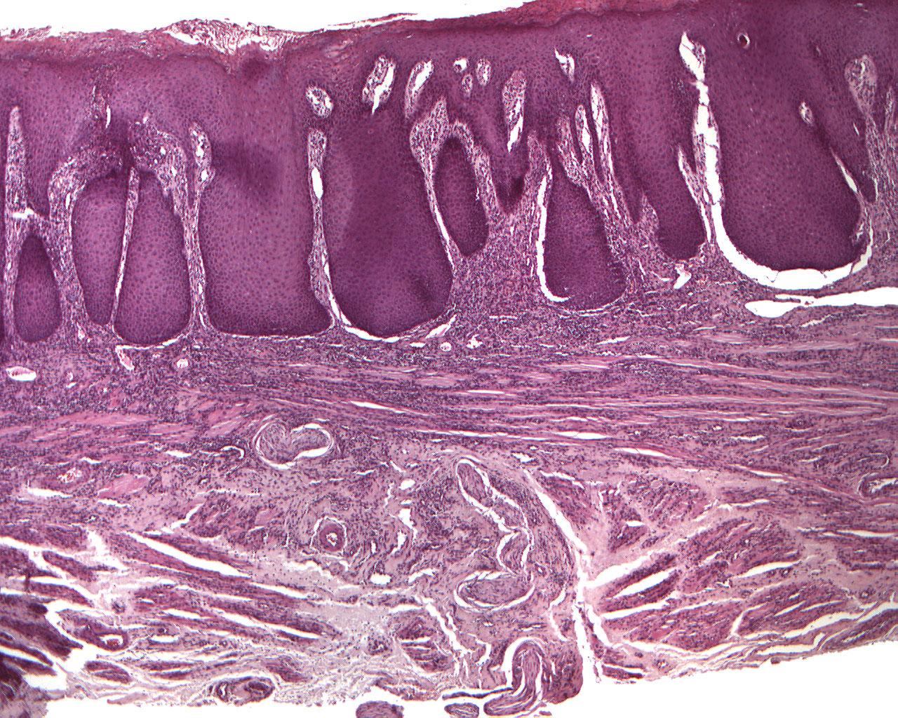
Fungal infected mucosa with squamous hyperplasia and moderate chronic inflammation (40X)
Clinical information: A 59-year-old man had a diffuse irritation/lesion on the left side of the dorsum of the tongue. The papillae were atrophic, but no induration (indurare, (lat.) = to make hard). The lesion was in direct contact with amalgam restorations. The whole lesion was excised.
Microscopic examination: Microscopically, the mucosal surface is covered by hyperparakeratinized, multi-layered squamous epithelium that exhibits considerable proliferation in depth, forming fairly wide ridges and networks. In some places micropustules containing mainly lymphocytes are found in the stratum corneum. A number of basal and suprabasal mitoses are seen in some of the epithelial outgrowths. The border against the underlying, loose connective tissue is well defined. The Underlying connectove tissueis diffusely infiltrated by a number of lymphocytes and plasma cells. The resection margin in deeper part of the tissue is limited by some striated musculature.
In the PAS colored section of the same tissue, you can see the hyphae of fungal infection.