

Bullous lichen ruber planus (40X)
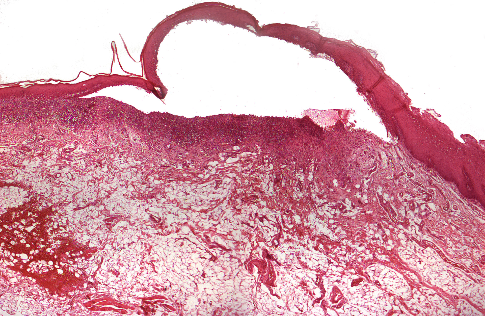
Clinical information: In a 74-year-old woman, white, lacy patches were incidentally detected. Painful bullae occasionally developed (Latin: bulla = bubble. Bladder. Used for blisters with a diameter of over 5 mm.)
Clinical diagnosis: Lichen planus
Microscopic examination: One can see mucous membrane covered by a multi-layered, hyperparakeratinized squamous epithelium. In some places, the epithelium is relatively thin and demarcated in a straight line from the underlying connective tissue - in other places it is thicker and tends to form a network (hyperplasia). In some areas, the basal cell layer is degeneratively changed with small blisters. Towards this part, the epithelium has clearly detached from the underlying connective tissue so that a bladder has appeared. The underlying connective tissue shows a "bed" of inflammatory cells, most of which are of a lymphocytic nature (T-lymphocytes). Deeper, the tissue is more fibrous and contains some fatty tissue.