

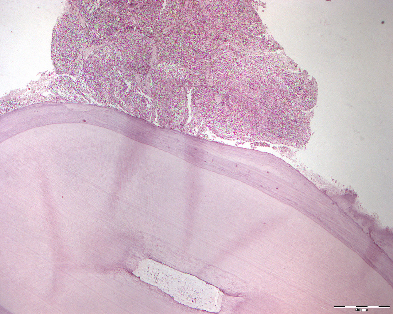
The apex of a root from a tooth with a granuloma containing squamous epithelial cells (40X)
Clinical information: A 42-year-old woman had diffuse, intermittent (L, inter=between; mitter=to send; interrupted, not continuous) pain in region of tooth 45. The tooth had a large mesial-occlusal amalgam filling. Radiologically, a small, well-defined clearing was present apically. The tooth was extracted.
Clinical diagnosis: ?
Microscopic examination: A root tip and a root canal is seen where in the root canal contins a loose, vascular, leukocyte-infiltrated tissue with small calcifications (vital pulp). On the surface and as a direct continuation of the root membrane, there is a granulation tissue wich copious amounts of cells and irregular connective tissue features. Small islands of epithelial tissue are seen aswell. In the largest islands, the epithelium tends to have a central decay with the formation of small cavities (microcysts). This is perceived as the first step towards cyst formation. Where do these epithelial islands come from? Why does the epithelium degenerate?