

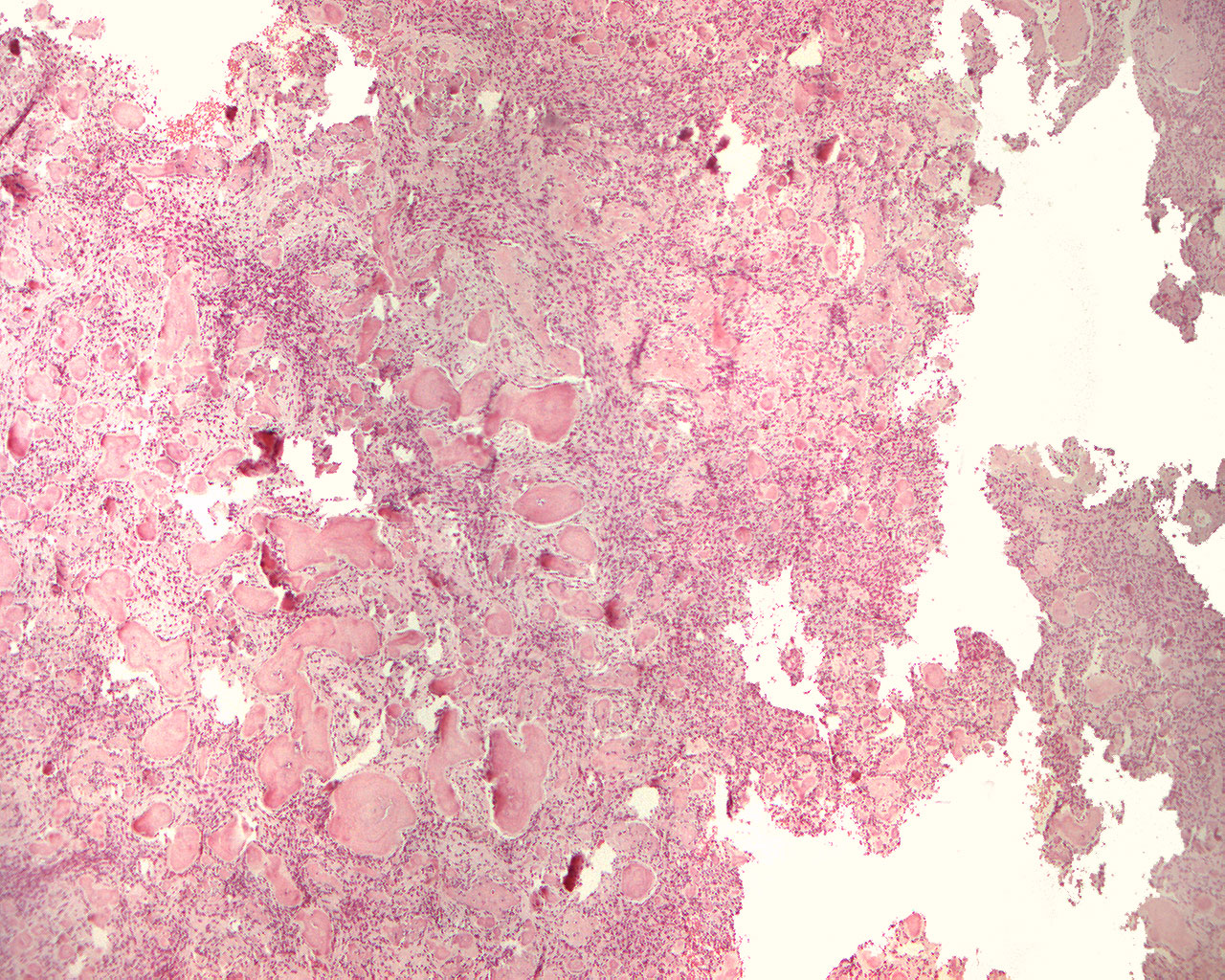
Cemento-ossifying fibroma (40X)
Clinical information: A 42-year-old woman had a missing tooth 34. In the area of the missing tooth, there was a tumor with sharp cortical detachment and expansion of the buccal and lingual cortical membrane. A homogeneous, cancellous bone mass with abundant vascularization was removed.
Clinical diagnosis: Odontogenic calcifying tumor.
Microscopic examination: Small tissue specimen are found, partly made up of irregular islands and beams of partly acellular, partly cellular hard tissue which looks like cement or bone. The hard tissue structures are covered by a cementoid (unmineralized) zone which is in turn covered in places by osteoblasts/cementoblasts. In between the hard tissue elements, a relatively fibroblast-rich connective tissue with thin-walled, partly blood-filled veins is found, but the vascularization is not strikingly large. However, copious amounts of blood clots are seen in in between the tissue specimen (not seen in this image).
Comment: Combined with the information that the process was radiologically well defined, the image is perceived as a cemento-ossifying fibroma. A similar histological picture can be seen in periapical cement dysplasia which, however, has diffuse radiological limitation.
There are no objective differences between cement and bone, but when hard tissue like this is seen in relation to a tooth, the term cemento-ossifying is often used.
Image caption: