

Follicular ameloblastoma (40X)
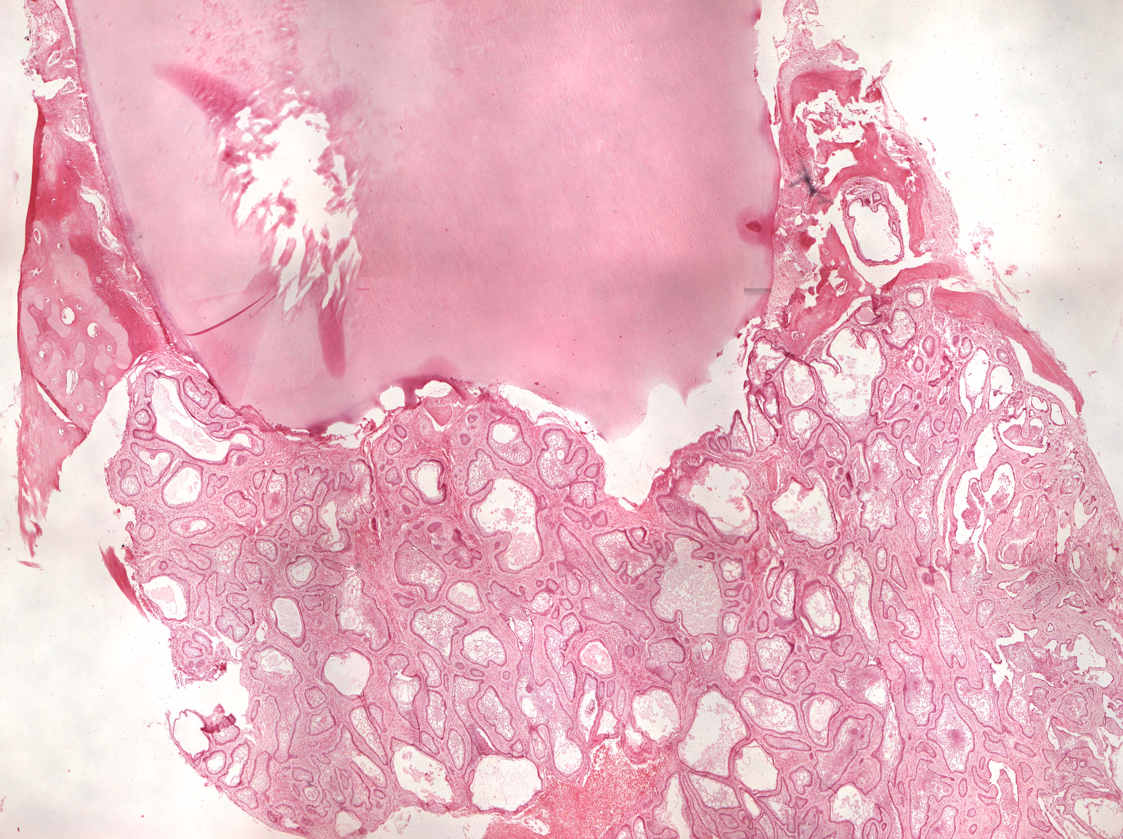
Clinical information: In a 64-year-old woman, a small buccal lesion / swelling close to teeth 33 and 34 was incidentally detected. Two years later, the swelling was the size of a nut buccally and could also be palpated lingually as a hard, solid tumor, without fluctuation and with moderate tenderness to applied pressure. Tooth 33 and 34 were vital (non-pulpogenic process!). The tumor together with adjacent alveolar bone as well as teeth 33 and 34 were removed.
Clinical diagnosis: Tumor mandibulae (ameloblastoma).
Microscopic examination: You can see a tooth where the pulp is not visible. Attached to the root surface, where there are some resorption defects in cementum and dentine, a non-encapsulated, epithelial tumor tissue is found made up of islands and flakes (follicles) of peripheral cylindrical, preameloblast-like cells and central round, oblong and star-shaped cells reminiscent of the star-shaped (stellate) reticulum (epithelial!). Some cystic decay is seen centrally in the epithelium (why?). In some areas there are som tendecy of epithelial keratinization. The surrounding connective tissue is relatively fibrous and does not show signs of being inductively changed (it is not cell-rich like the dental papilla). To the sides, some compact bone tissue with ingrowth of tumor tissue is seen.
Comment: The ameloblastoma is a benign, but locally aggressive, epithelial, odontogenic tumor without odontogenic mesenchyme. It is therefore relatively poorly differentiated compared to normal odontogenesis (consists only of enamel organ-like tissue). (Differentiation, from Latin dis = apart; ferre = carry, lead: development in different directions; structural and/or functional modification of tissues or organs during development).