

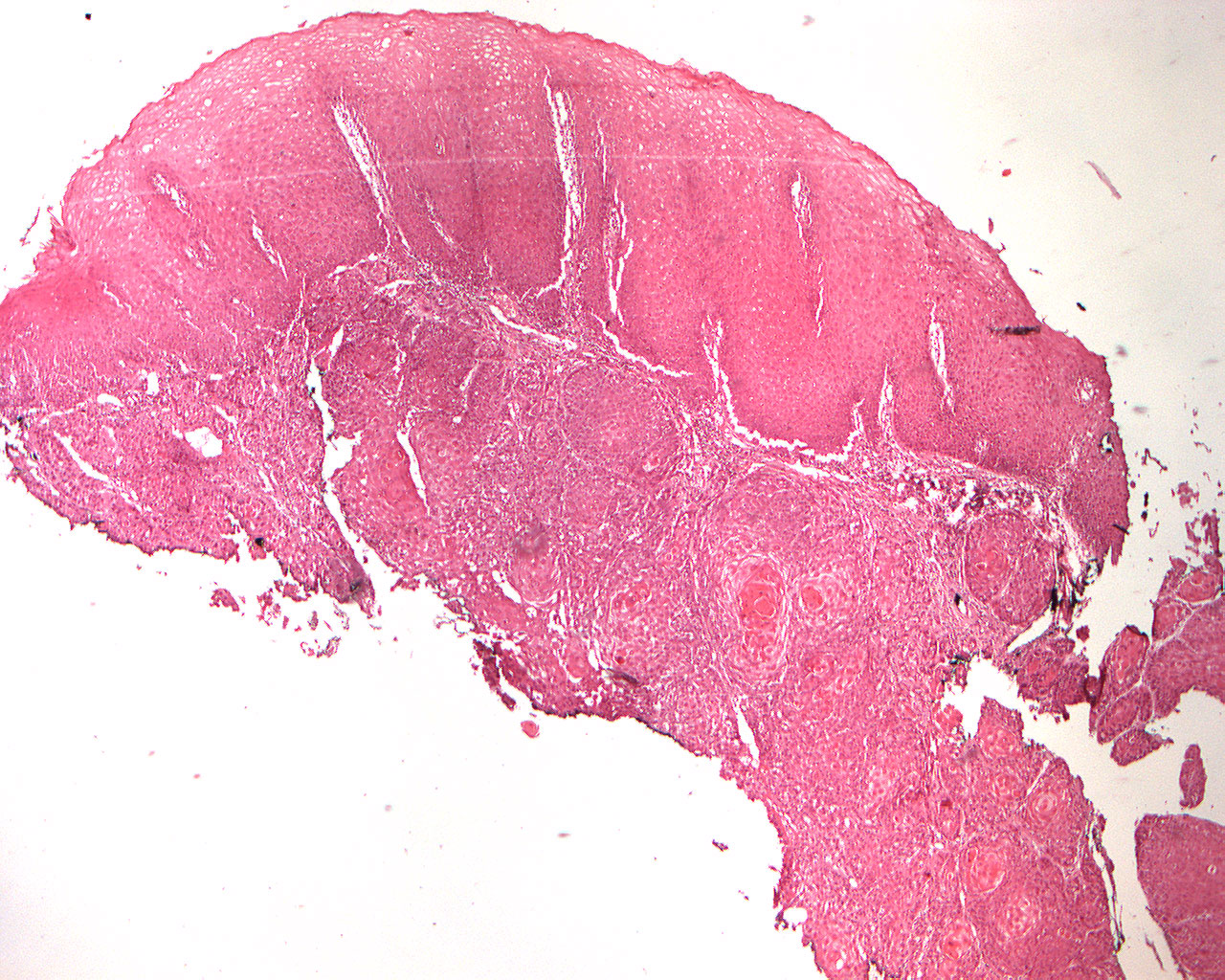
Squamous cell carcinoma (40X)
Clinical information: There was ulcerating denture bite lingual region 46, 47 in a 69-year-old man. The condition was suspicious for malignancy.
Clinical diagnosis: cancer of the mouth
Microscopic examination: one can see pieces of mucosa covered by a multilayered squamous epithelium with a partly parakeratinized, partly hyperparakeratinized surface and partly thick stratum spinosum and multiplied basal cell layer (basilar hyperplasia)- There is some variation in the shape, size and coloring of the cells (cellular polymorphism) and also in the size, shape and coloring of the nuclei (nuclear polymorphism). On one side of the specimen, the dysplastic epithelial covering turns into an infiltrating tumor tissue with islands, strings, flakes and streaks of epithelial cells where the cellular and nuclear changes described above are more pronounced. Significant keratinization of individual cells as well as of groups of cells (onion formation, horn pearl formation) is found. The connective tissue shows diffuse, moderate to intense infiltration of mononuclear inflammatory cells.
Comment: There is no evidence of a traumatic etiology (e.g. denture bite) in cancer, but trauma can aggravate the condition. If alleviation of gnawing (prosthesis, tooth/filling edge) does not improve within 14 days, a change such as here should be considered cancer until the opposite is proven and a biopsy shoud be carried out.