

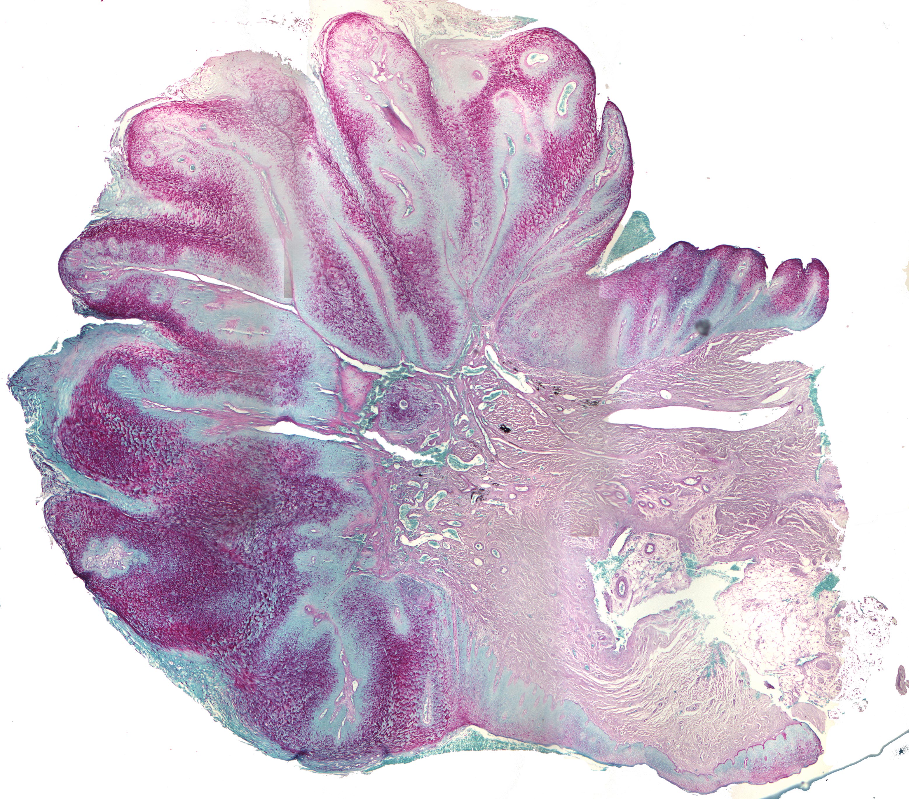
Fungal infection of a verrucous carcinoma (PAS) (40X)
Clinical information: See section the H+E stained section of the fungal infected verrucous carcinoma as the tissue is from the same preparation.
Clinical diagnosis: Exophytic tumor
Microscopic examination: This specially stained preparation (PAS-CYT) shows abundant occurrence of hyphae and pseudohyphae compatible with the pathogenic form of Candida albicans growing in the parakeratin layer. The fungal hyphae appear as purple-coloured, straight or slightly curved threads mostly perpendicular to the tissue surface. Unlike hyphae, pseudohyphae have cytoplasmic constrictions.
Comment: PAS staining gives a purple-red reaction with aldehyde groups, possibly after opening of ring structures (unmasking). A positive reaction is therefore seen in the wall of the hyphae/pseudohyphae, which is made up of cellulose, a carbohydrate consisting of hexoses. The large areas of fine granular, red substance represent keratohyalin and have nothing to do with the fungal infection.