

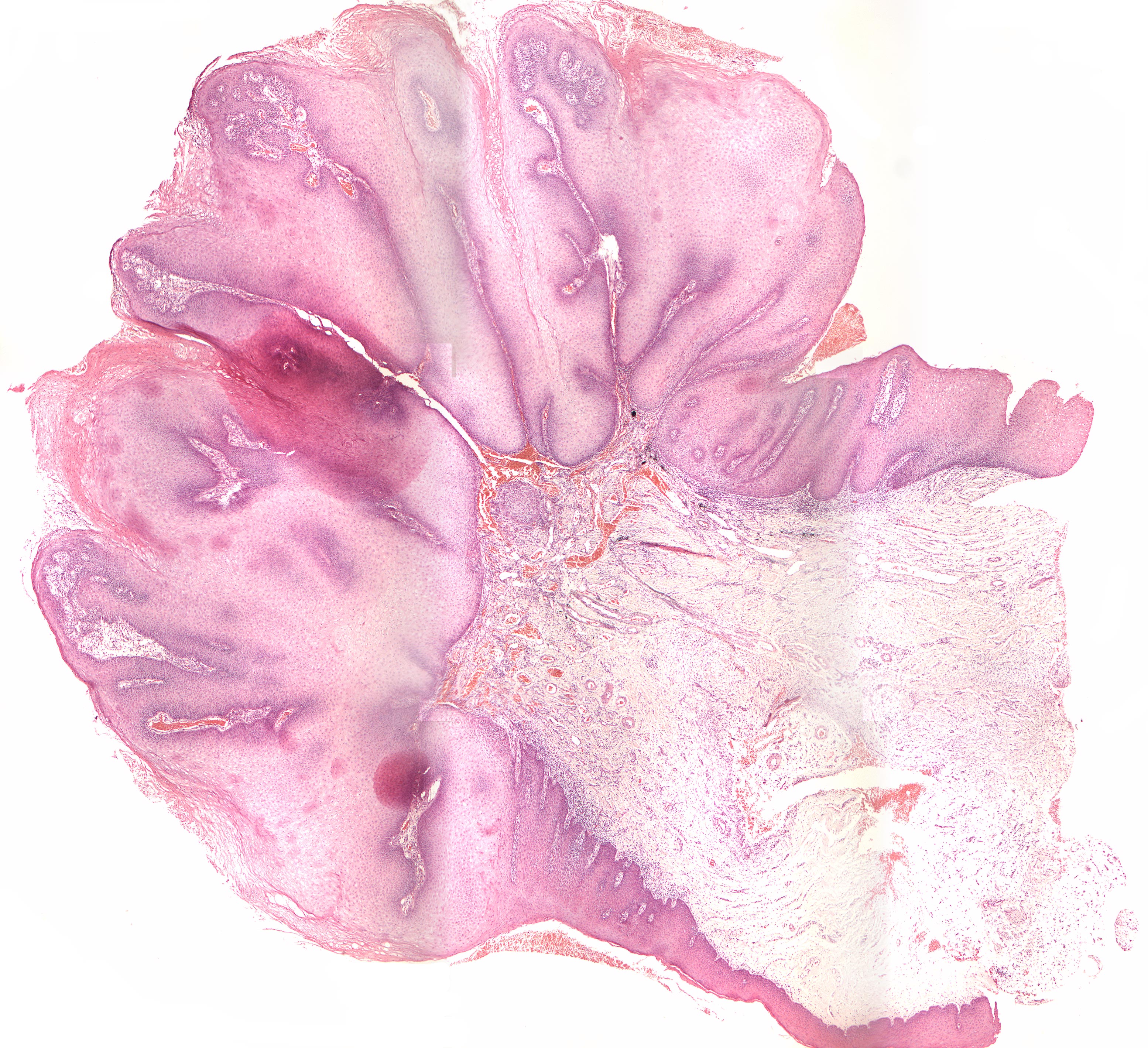
Fungal infection of a verrucous carcinoma (H+E) (40X)
Clinical information: In a 72-year-old man, a leukoplakic lesion close to tooth 37 had been extirpated about 14 years earlier. Now he presented himself with with a round, exophytic (Latin: exo= out; Greek: phyllon= leaf; growing outwards like a plant) 0.5 x 0.5 cm whitish, verrucous tumor in the same region. The preparation originates from here. The patient also had a slightly elevated, whitish, verrucous, 1 x 0.3 cm tumor regio 47.
Clinical diagnosis: Exophytic tumor
Microscopic examination: Microscopically, broad-stalked mucosal preparations are seen whose strongly convex surface is covered by multi-layered squamous epithelium with significant hyperparakeratosis and the presence of parakeratin-filled crypts. The epithelium proliferates in depth with the the formation of a broad-beamed network and close-set, wide spikes ("elephant feet") with a pushing front towards the connective tissue. The stratum spinosum appears regularly and inconspicuously over large areas, but in some places there is some variation in the size, shape and coloration of the cells. Numerous large mitosis figures are also seen high in the stratum spinosum and down towards the basal cell layer. This occurs in large areas with several shoal-like layers (basilar hyperplasia) and produces some variation in the size, shape and colorability of cells and nuclei (cellular and nuclear polymorphism). The border close to the underlying loose connective tissue is well defined. There are small island of tumor tissue within the connective tissue, but otherwise no definite infiltrating growth. The connective tissue is loosely structured and presents numerous hyperemic, dilated veins and diffuse, mild to moderate infiltration of lymphocytes and some plasma cells. As a secondary finding, some fine granular and herniated black foreign matter compatible with amalgam components is seen. Down in the stem of the specimen, connective tissue with varying degrees of mononuclear infiltration is found.
Comment: Typical of the verrucous carcinoma is its exophytic character and pushing (non-infiltrating) limitation towards the connective tissue, the hyperparakeratosis and in particular the parakeratin-filled crypts as well as the tumor tissue's relatively high degree of differentiation (slight to moderate polymorphism) with relatively good stratification and often only very few mitoses.