

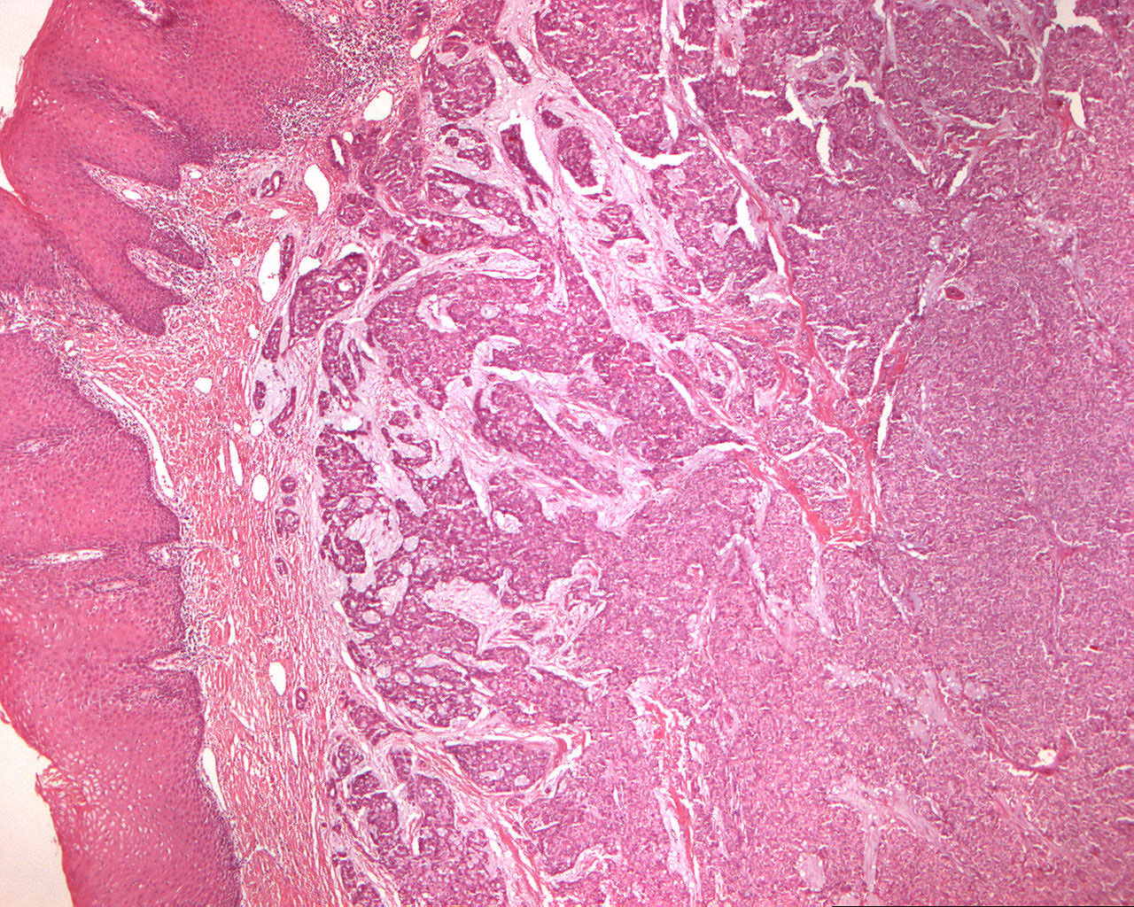
Adenocystic carcinoma of the palate (40X)
Clinical information: A 50-year-old woman had an approx. plum-sized tumor in the hard palate. The tissue was obtained from the same patient as in this section.
Clinical diagnosis: Palatinal tumor
Microscopic examination: The tumor tissue shows several morphological patterns; you can partly see flakes, strings and tubular structures consisting of isomorphic (iso. Greek = equal) epithelial cells interspersed with some lighter cells with dark nuclei, partly you can see areas where the tumor tissue has the typical adenocystic appearance with tumor islands of different sizes perforated by small and larger holes (cribriform = sieve-like pattern; cribrum(L) = sieve). The stroma is a loose connective tissue that appears slightly mucinous in some places. The tumor is not encapsulated and grows infiltratively in the surroundings; locally it "attacks" the epithelial surface.